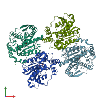
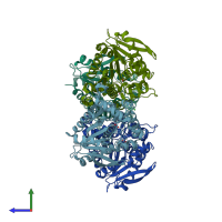
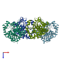
5jd5

X-ray diffraction
1.95Å resolution

Crystal structure of MGS-MilE3, an alpha/beta hydrolase enzyme from the metagenome of pyrene-phenanthrene enrichment culture with sediment sample of Milazzo Harbor, Italy

Released:
Source organism: uncultured bacterium
Entry authors: Stogios PJ, Xu X, Cui H, Martinez-Martinez M, Chernikova TN, Golyshin PN, Yakimov MM, Ferrer M, Savchenko A

Function and Biology Details

Biochemical function:
Biological process:
  • not assigned
Cellular component:
  • not assigned

Structure analysis Details

Assemblies composition:
monomeric (preferred)
homo dimer
Assembly name:
PDBe Complex ID:
PDB-CPX-165862 (preferred)
Entry contents:
1 distinct polypeptide molecule
Macromolecule:
MGS-MilE3 Chains: A, B, C, D
Molecule details ›
Chains: A, B, C, D
Length: 321 amino acids
Theoretical weight: 34.81 KDa
Source organism: uncultured bacterium
Expression system: Escherichia coli BL21(DE3)
Structure domains: alpha/beta hydrolase

Ligands and Environments

1 bound ligand:
1 modified residue:

Experiments and Validation Details

Entry percentile scores
X-ray source: RIGAKU MICROMAX-007 HF
Spacegroup: P212121
Unit cell:
a: 91.668Å b: 97.16Å c: 167.67Å
α: 90° β: 90° γ: 90°
R-values:
R R work R free
0.196 0.195 0.237
Expression system: Escherichia coli BL21(DE3)