


Function and Biology Details
Reaction catalysed:
2 glutathione + ROOH = glutathione disulfide + H(2)O + ROH
Biochemical function:
Biological process:
Cellular component:
Sequence domains:
Structure analysis Details
Assembly composition:
homo dimer (preferred)
Assembly name:
Glutathione-dependent peroxiredoxin (preferred)
PDBe Complex ID:
PDB-CPX-103905 (preferred)
Entry contents:
1 distinct polypeptide molecule
Macromolecule:
Glutathione-dependent peroxiredoxin
Molecule details ›
Chain: A
Length: 164 amino acids
Theoretical weight: 17.55 KDa
Source organism: Vibrio vulnificus MO6-24/O
Expression system: Escherichia coli
UniProt:
Structure domains: Glutaredoxin
Length: 164 amino acids
Theoretical weight: 17.55 KDa
Source organism: Vibrio vulnificus MO6-24/O
Expression system: Escherichia coli
UniProt:
- Canonical:
 A0A1Z0YU25 (Residues: 1-164; Coverage: 100%)
Structure domains: Glutaredoxin
Ligands and Environments
No bound ligands
No modified residues
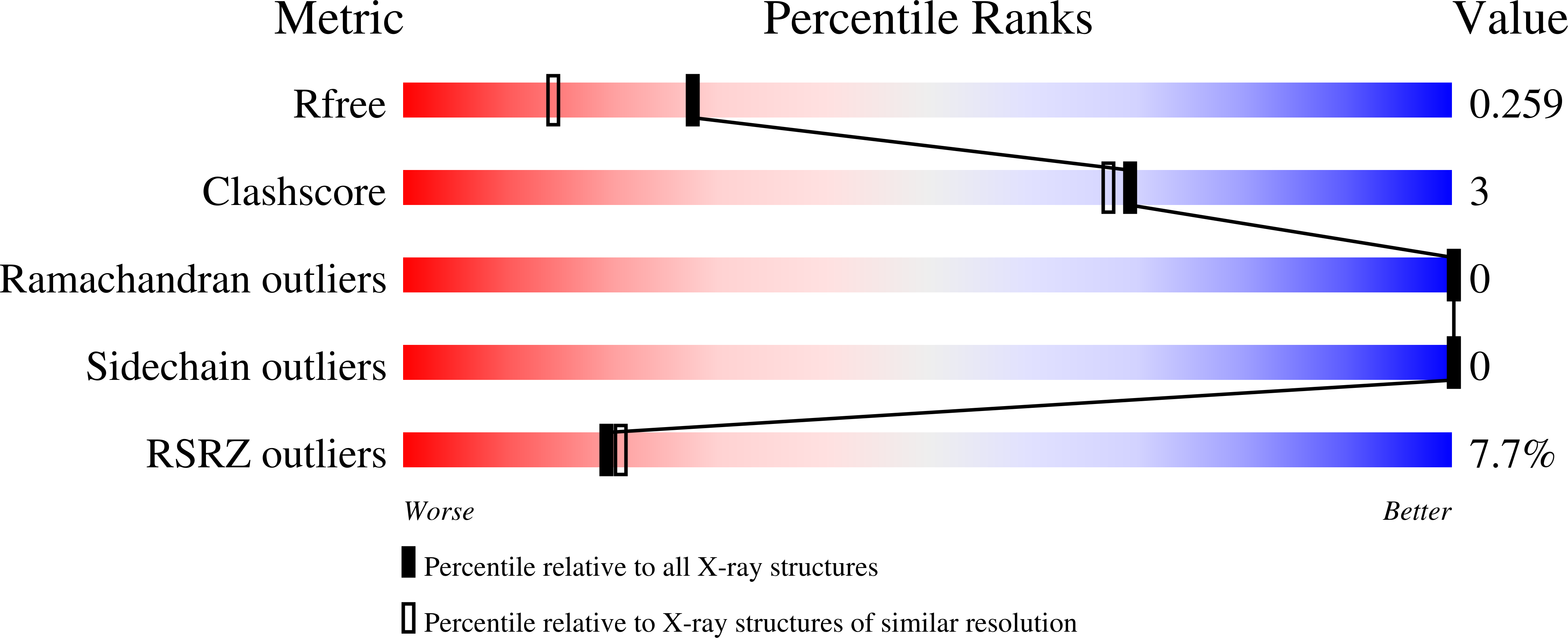
Experiments and Validation Details
wwPDB Validation report is not available for this entry.
X-ray source:
PAL/PLS BEAMLINE 5C (4A)
Spacegroup:
P3221
Expression system: Escherichia coli
