5kkp

X-ray diffraction
2.26Å resolution

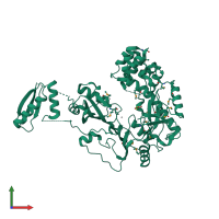
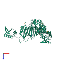
Crystal Structure of Human Pseudouridylate Synthase 7

Released:
Source organism: Homo sapiens
Entry authors: DONG A, ZENG H, WALKER JR, Bountra C, Arrowsmith CH, Edwards AM, BROWN PJ, WU H, Structural Genomics Consortium (SGC)

Function and Biology Details

Structure analysis Details

Assembly composition:
monomeric (preferred)
PDBe Complex ID:
PDB-CPX-188685 (preferred)
Entry contents:
1 distinct polypeptide molecule
Macromolecule:
Pseudouridylate synthase 7 homolog Chain: A
Molecule details ›
Chain: A
Length: 563 amino acids
Theoretical weight: 65.25 KDa
Source organism: Homo sapiens
Expression system: Escherichia coli BL21(DE3)
UniProt:
  • Canonical: Q96PZ0 (Residues: 99-661; Coverage: 85%)
Gene names: KIAA1897, PUS7
Sequence domains: tRNA pseudouridine synthase D (TruD)

Ligands and Environments

2 bound ligands:
1 modified residue:

Experiments and Validation Details

Entry percentile scores
X-ray source: APS BEAMLINE 22-ID
Spacegroup: P212121
Unit cell:
a: 56.356Å b: 73.4Å c: 138.179Å
α: 90° β: 90° γ: 90°
R-values:
R R work R free
0.21 0.209 0.263
Expression system: Escherichia coli BL21(DE3)