5l03

X-ray diffraction
1.47Å resolution

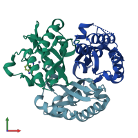
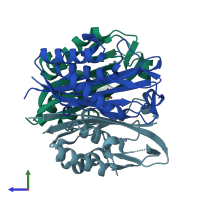
Crystal structure of 2-C-METHYL-D-ERYTHRITOL 2,4-CYCLODIPHOSPHATE Synthase from BURKHOLDERIA PSEUDOMALLEI bound to L-tryptophan hydroxamate

Released:
Source organism: Burkholderia pseudomallei
Entry authors: Blain JM, Ghose D, Gorman JL, Goshu GM, Ranieri G, Zhao L, Bode B, Meganathan R, Walter RL, Hagen TJ, Horn JR

Function and Biology Details

Reaction catalysed:
2-phospho-4-(cytidine 5'-diphospho)-2-C-methyl-D-erythritol = 2-C-methyl-D-erythritol 2,4-cyclodiphosphate + CMP
Biochemical function:
Cellular component:
  • not assigned

Structure analysis Details

Assembly composition:
homo trimer (preferred)
PDBe Complex ID:
PDB-CPX-100409 (preferred)
Entry contents:
1 distinct polypeptide molecule
Macromolecule:
2-C-methyl-D-erythritol 2,4-cyclodiphosphate synthase Chains: A, B, C
Molecule details ›
Chains: A, B, C
Length: 158 amino acids
Theoretical weight: 16.86 KDa
Source organism: Burkholderia pseudomallei
Expression system: Escherichia coli
UniProt:
  • Canonical: A0A069B2G5 (Residues: 1-158; Coverage: 98%)
Gene names: Y036_3499, ispF
Sequence domains: YgbB family

Ligands and Environments

2 bound ligands:
No modified residues

Experiments and Validation Details

Entry percentile scores
X-ray source: APS BEAMLINE 22-BM
Spacegroup: C2
Unit cell:
a: 116.515Å b: 68.126Å c: 60.659Å
α: 90° β: 96.93° γ: 90°
R-values:
R R work R free
0.18 0.179 0.2
Expression system: Escherichia coli