We've built a new version of this page with an interactive 3D viewer. Try it out in beta and help us improve:

5lqz

Electron Microscopy
7Å resolution

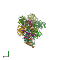
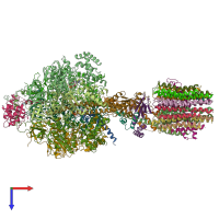
Structure of F-ATPase from Pichia angusta, state1

Released:
Source organism: Ogataea angusta
Related structures: EMD-4102

Function and Biology Details

Structure analysis Details

Assembly composition:
hetero 30-mer (preferred)
PDBe Complex ID:
PDB-CPX-123063 (preferred)
Entry contents:
17 distinct polypeptide molecules
Macromolecules (17 distinct):
ATP synthase subunit f Chain: 1
Molecule details ›
Chain: 1
Length: 30 amino acids
Theoretical weight: 2.57 KDa
Source organism: Ogataea angusta
ATP synthase subunit AAP1 Chain: 2
Molecule details ›
Chain: 2
Length: 25 amino acids
Theoretical weight: 2.15 KDa
Source organism: Ogataea angusta
ATP synthase subunit a Chain: 3
ATP synthase subunit b Chain: 4
ATP synthase subunit alpha Chains: A, B, C
ATP synthase subunit beta Chains: D, E, F
ATP synthase subunit gamma Chain: G
ATP synthase subunit delta, mitochondrial Chain: H
ATP synthase epsilon subunit Chain: I
ATPase inhibitor, mitochondrial Chain: J
ATP synthase subunit 9, mitochondrial Chains: K, L, M, N, O, P, Q, R, S, T
ATP synthase subunit 5, mitochondrial Chain: U
ATP synthase subunit 4 Chain: V
ATP synthase subunit d, mitochondrial Chain: W
ATP synthase subunit h Chain: X
ATP synthase subunit a Chain: Y
ATP synthase subunit a Chain: Z

Ligands and Environments

3 bound ligands:
No modified residues

Experiments and Validation Details

wwPDB Validation report is not available for this EM entry.
Resolution:
Relevant EMDB volumes: EMD-4102