

methyl]-~{N}1-(cyclohexylmethyl)-~{N}4-cyclopentyl-~{N}1-[(~{Z})-4-[(~{E})-methyliminomethyl]-5-oxidanyl-hex-4-enyl]benzene-1,4-disulfonamide</span>;</li> <li class='image_legend_li'>1 copy of <span class='highlight'>water</span>.</li></ul>")
methyl]-~{N}1-(cyclohexylmethyl)-~{N}4-cyclopentyl-~{N}1-[(~{Z})-4-[(~{E})-methyliminomethyl]-5-oxidanyl-hex-4-enyl]benzene-1,4-disulfonamide</span>;</li> <li class='image_legend_li'>1 copy of <span class='highlight'>water</span>.</li></ul>")
methyl]-~{N}1-(cyclohexylmethyl)-~{N}4-cyclopentyl-~{N}1-[(~{Z})-4-[(~{E})-methyliminomethyl]-5-oxidanyl-hex-4-enyl]benzene-1,4-disulfonamide</span>;</li> <li class='image_legend_li'>1 copy of <span class='highlight'>water</span>.</li></ul>")
Function and Biology Details
Biochemical function:
Biological process:
Cellular component:
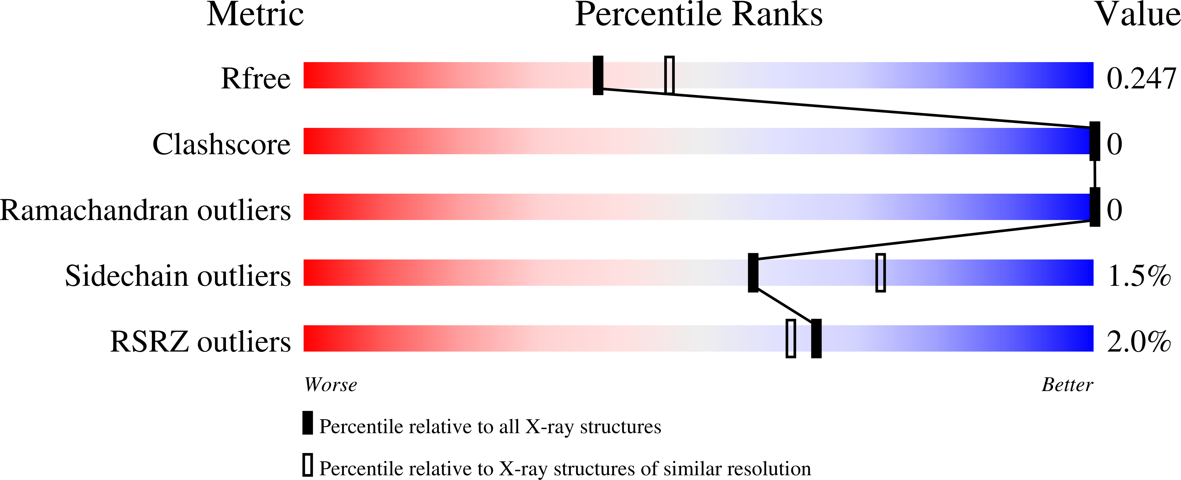
Structure analysis Details
Assembly composition:
monomeric (preferred)
Assembly name:
PDBe Complex ID:
PDB-CPX-129171 (preferred)
Entry contents:
1 distinct polypeptide molecule
Macromolecule:
Retinal rod rhodopsin-sensitive cGMP 3',5'-cyclic phosphodiesterase subunit delta
Molecule details ›
Chain: B
Length: 150 amino acids
Theoretical weight: 17.44 KDa
Source organism: Homo sapiens
Expression system: Escherichia coli
UniProt:
Sequence domains: GMP-PDE, delta subunit
Structure domains: GMP phosphodiesterase, delta subunit
Length: 150 amino acids
Theoretical weight: 17.44 KDa
Source organism: Homo sapiens
Expression system: Escherichia coli
UniProt:
- Canonical:
 O43924 (Residues: 1-150; Coverage: 100%)
Sequence domains: GMP-PDE, delta subunit
Structure domains: GMP phosphodiesterase, delta subunit

