


Function and Biology Details
Reaction catalysed:
Hydrolysis of (1->4)-beta-linkages between N-acetylmuramic acid and N-acetyl-D-glucosamine residues in a peptidoglycan and between N-acetyl-D-glucosamine residues in chitodextrins
Biochemical function:
Biological process:
Cellular component:
Structure analysis Details
Assembly composition:
monomeric (preferred)
Assembly name:
Endolysin (preferred)
PDBe Complex ID:
PDB-CPX-112953 (preferred)
Entry contents:
1 distinct polypeptide molecule
Macromolecule:
Endolysin
Molecule details ›
Chain: A
Length: 164 amino acids
Theoretical weight: 18.58 KDa
Source organism: Escherichia virus T4
Expression system: Escherichia coli BL21
UniProt:
Sequence domains: Phage lysozyme
Length: 164 amino acids
Theoretical weight: 18.58 KDa
Source organism: Escherichia virus T4
Expression system: Escherichia coli BL21
UniProt:
- Canonical:
 D9IEF7 (Residues: 1-164; Coverage: 100%)
Sequence domains: Phage lysozyme
Ligands and Environments
No bound ligands
No modified residues
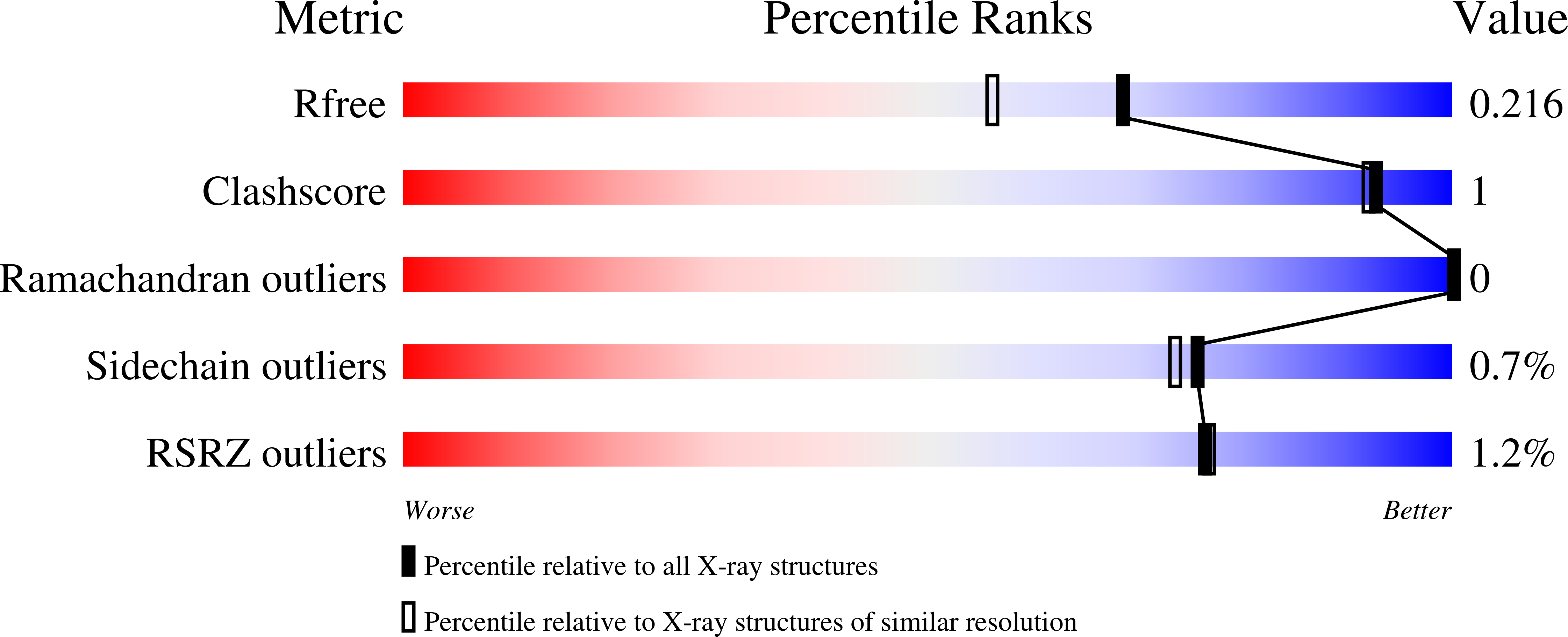
Experiments and Validation Details
wwPDB Validation report is not available for this entry.
X-ray source:
RIGAKU RUH2R
Spacegroup:
P3221
Expression system: Escherichia coli BL21
