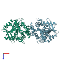
5o2g

X-ray diffraction
2.1Å resolution

Crystal structure determination from picosecond infrared laser ablated protein crystals by serial synchrotron crystallography

Released:
Source organism: Rhodopseudomonas palustris
Entry authors: Schulz EC, Kaub J, Busse F, Mehrabi P, Mueller-Werkmeiser H, Pai EF, Robertson WD, Miller RJD

Function and Biology Details

Reaction catalysed:
Haloacetate + H(2)O = glycolate + halide
Biochemical function:
Biological process:
  • not assigned
Cellular component:
  • not assigned

Structure analysis Details

Assembly composition:
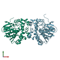
homo dimer (preferred)
PDBe Complex ID:
PDB-CPX-104436 (preferred)
Entry contents:
1 distinct polypeptide molecule
Macromolecule:
AB hydrolase-1 domain-containing protein Chains: A, B
Molecule details ›
Chains: A, B
Length: 306 amino acids
Theoretical weight: 34.07 KDa
Source organism: Rhodopseudomonas palustris
Expression system: Escherichia coli
UniProt:
  • Canonical: A0A2R4GQN1 (Residues: 1-302; Coverage: 100%)
Gene name: RPYSC3_11920
Sequence domains: alpha/beta hydrolase fold

Ligands and Environments

No bound ligands
No modified residues

Experiments and Validation Details

Entry percentile scores
X-ray source: PETRA III, EMBL c/o DESY BEAMLINE P14 (MX2)
Spacegroup: P21
Unit cell:
a: 42.413Å b: 80.349Å c: 85.648Å
α: 90° β: 102.96° γ: 90°
R-values:
R R work R free
0.225 0.222 0.27
Expression system: Escherichia coli