


Function and Biology Details
Biochemical function:
- not assigned
Biological process:
- not assigned
Cellular component:
- not assigned
Structure analysis Details
Assembly composition:
monomeric (preferred)
Assembly name:
PDBe Complex ID:
PDB-CPX-191188 (preferred)
Entry contents:
1 distinct polypeptide molecule
Macromolecule:
Glycoside-hydrolase family GH114 TIM-barrel domain-containing protein
Molecule details ›
Chain: A
Length: 278 amino acids
Theoretical weight: 30.52 KDa
Source organism: Pseudomonas aeruginosa PAO1
Expression system: Escherichia coli
UniProt:
Sequence domains: Glycoside-hydrolase family GH114
Length: 278 amino acids
Theoretical weight: 30.52 KDa
Source organism: Pseudomonas aeruginosa PAO1
Expression system: Escherichia coli
UniProt:
- Canonical:
 Q9HZE4 (Residues: 47-303; Coverage: 27%)
Sequence domains: Glycoside-hydrolase family GH114
Ligands and Environments
No bound ligands
No modified residues
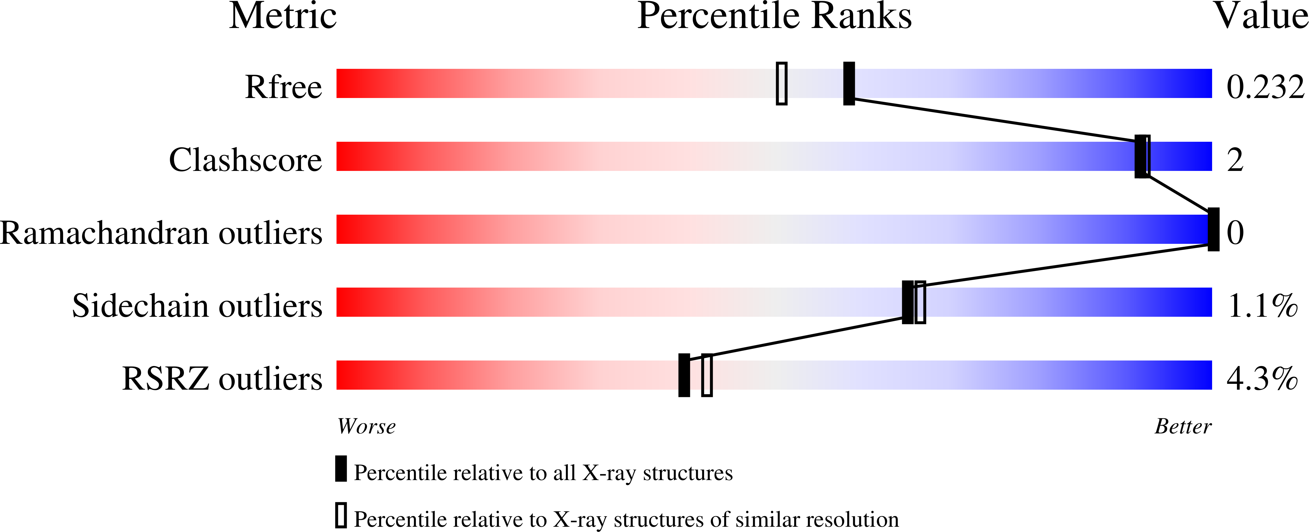
Experiments and Validation Details
wwPDB Validation report is not available for this entry.
X-ray source:
BRUKER D8 QUEST
Spacegroup:
P21212
Expression system: Escherichia coli
