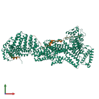
5u1t

X-ray diffraction
2.6Å resolution

Crystal structure of the Saccharomyces cerevisiae separase-securin complex at 2.6 angstrom resolution

Released:
Primary publication:
Molecular mechanism for the regulation of yeast separase by securin.
Nature 542 255-259 (2017)
PMID: 28146474

Function and Biology Details

Reaction catalysed:
All bonds known to be hydrolyzed by this endopeptidase have arginine in P1 and an acidic residue in P4. P6 is often occupied by an acidic residue or by a hydroxy-amino-acid residue, the phosphorylation of which enhances cleavage.
Biochemical function:
Biological process:
Cellular component:

Structure analysis Details

Assembly composition:
hetero dimer (preferred)
PDBe Complex ID:
PDB-CPX-154072 (preferred)
Entry contents:
2 distinct polypeptide molecules
Macromolecules (2 distinct):
Separin Chain: A
Molecule details ›
Chain: A
Length: 1596 amino acids
Theoretical weight: 184.04 KDa
Source organism: Saccharomyces cerevisiae S288C
Expression system: Trichoplusia ni
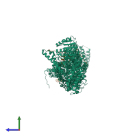
UniProt:
  • Canonical: Q03018 (Residues: 51-1630; Coverage: 97%)
Gene names: ESP1, YGR098C
Sequence domains: Peptidase family C50
Securin Chain: B
Molecule details ›
Chain: B
Length: 117 amino acids
Theoretical weight: 13.37 KDa
Source organism: Saccharomyces cerevisiae S288C
Expression system: Trichoplusia ni
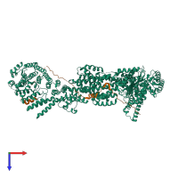
UniProt:
  • Canonical: P40316 (Residues: 257-373; Coverage: 31%)
Gene names: PDS1, YD9727.08C, YDR113C
Sequence domains: Securin sister-chromatid separation inhibitor

Ligands and Environments

No bound ligands
No modified residues

Experiments and Validation Details

Entry percentile scores
X-ray source: APS BEAMLINE 24-ID-C
Spacegroup: P3221
Unit cell:
a: 125.863Å b: 125.863Å c: 271.944Å
α: 90° β: 90° γ: 120°
R-values:
R R work R free
0.222 0.221 0.264
Expression system: Trichoplusia ni