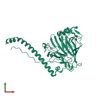
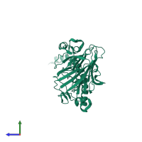
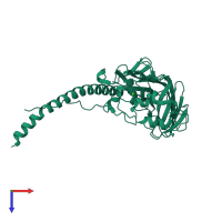
5uqz

X-ray diffraction
1.15Å resolution

Structural Analysis of the Glucan Binding Protein C of Streptococcus mutans Provides Evidence that it Mediates both Sucrose-Independent and -Dependent Adherence

Released:

Function and Biology Details

Biochemical function:
  • not assigned
Biological process:
  • not assigned
Cellular component:
  • not assigned

Structure analysis Details

Assemblies composition:
monomeric (preferred)
homo dimer
PDBe Complex ID:
PDB-CPX-184198 (preferred)
Entry contents:
1 distinct polypeptide molecule
Macromolecule:
Gram-positive cocci surface proteins LPxTG domain-containing protein Chain: A
Molecule details ›
Chain: A
Length: 354 amino acids
Theoretical weight: 39.25 KDa
Source organism: Streptococcus mutans UA159
Expression system: Escherichia coli BL21(DE3)
UniProt:
  • Canonical: Q8DTF1 (Residues: 124-477; Coverage: 64%)
Gene names: SMU_1396, gbpC
Sequence domains: Glucan-binding protein C

Ligands and Environments

1 bound ligand:
No modified residues

Experiments and Validation Details

Entry percentile scores
X-ray source: APS BEAMLINE 24-ID-E
Spacegroup: C2
Unit cell:
a: 213.098Å b: 48.596Å c: 40.775Å
α: 90° β: 96.29° γ: 90°
R-values:
R R work R free
0.126 0.124 0.146
Expression system: Escherichia coli BL21(DE3)