5xbf

X-ray diffraction
1.8Å resolution

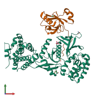
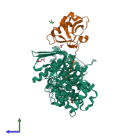
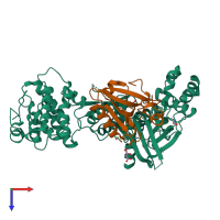
Crystal Structure of Myo7b C-terminal MyTH4-FERM in complex with USH1C PDZ3

Released:
Source organism: Homo sapiens
Primary publication:
Structure of Myo7b/USH1C complex suggests a general PDZ domain binding mode by MyTH4-FERM myosins.
Proc Natl Acad Sci U S A 114 E3776-E3785 (2017)
PMID: 28439001

Function and Biology Details

Structure analysis Details

Assembly composition:
hetero dimer (preferred)
PDBe Complex ID:
PDB-CPX-180168 (preferred)
Entry contents:
2 distinct polypeptide molecules
Macromolecules (2 distinct):
Unconventional myosin-VIIb Chain: A
Molecule details ›
Chain: A
Length: 516 amino acids
Theoretical weight: 59.44 KDa
Source organism: Homo sapiens
Expression system: Escherichia coli
UniProt:
  • Canonical: Q6PIF6 (Residues: 1601-2116; Coverage: 24%)
Gene name: MYO7B
Sequence domains:
Harmonin Chain: B
Molecule details ›
Chain: B
Length: 125 amino acids
Theoretical weight: 13.86 KDa
Source organism: Homo sapiens
Expression system: Escherichia coli
UniProt:
  • Canonical: Q9Y6N9 (Residues: 428-552; Coverage: 23%)
Gene names: AIE75, USH1C
Sequence domains: PDZ domain
Structure domains: Pdz3 Domain

Ligands and Environments

3 bound ligands:
No modified residues

Experiments and Validation Details

Entry percentile scores
X-ray source: SSRF BEAMLINE BL17U
Spacegroup: P21
Unit cell:
a: 69.634Å b: 42.537Å c: 118.39Å
α: 90° β: 97.51° γ: 90°
R-values:
R R work R free
0.188 0.187 0.211
Expression system: Escherichia coli