


Function and Biology Details
Reaction catalysed:
4 Fe(2+) + 4 H(+) + O(2) = 4 Fe(3+) + 2 H(2)O
Biochemical function:
Biological process:
Cellular component:
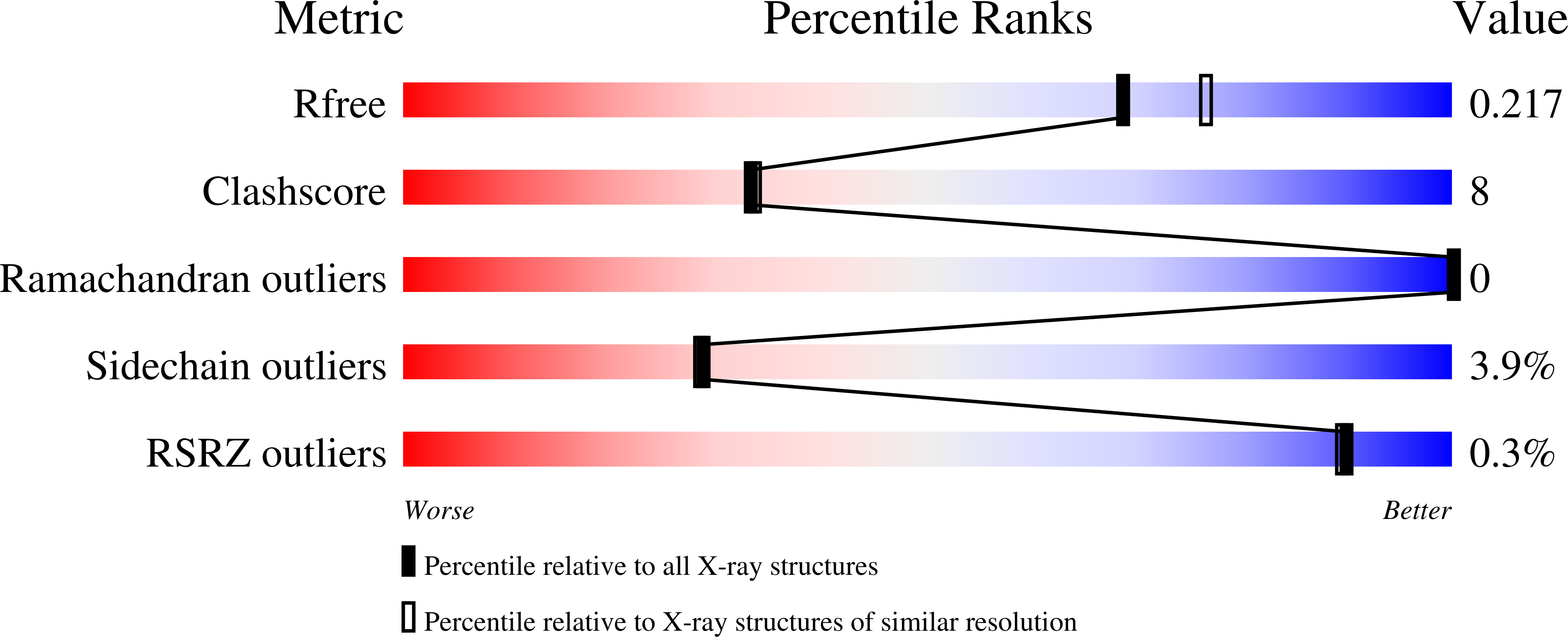
Structure analysis Details
Assembly composition:
homo dodecamer (preferred)
Assembly name:
PDBe Complex ID:
PDB-CPX-142037 (preferred)
Entry contents:
1 distinct polypeptide molecule
Macromolecule:
Bacterioferritin; DNA protection during starvation protein
Molecule details ›
Chains: A, B, C, D, E, F, G, H, I, J, K, L
Length: 200 amino acids
Theoretical weight: 22.66 KDa
Source organism: Escherichia coli K-12
Expression system: Escherichia coli
UniProt:
Sequence domains: Ferritin-like domain
Structure domains: Ferritin
Length: 200 amino acids
Theoretical weight: 22.66 KDa
Source organism: Escherichia coli K-12
Expression system: Escherichia coli
UniProt:
- Canonical:
 P0ABT2 (Residues: 1-163; Coverage: 98%)
 - Canonical: P0ABD3 (Residues: 138-158; Coverage: 13%)
Sequence domains: Ferritin-like domain
Structure domains: Ferritin

