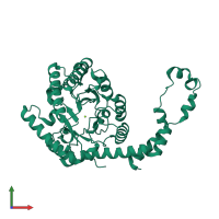
5y4j

X-ray diffraction
1.4Å resolution

Crystal structure of glucose isomerase in complex with xylitol inhibitor in one metal binding mode

Released:
Source organism: Streptomyces rubiginosus
Primary publication:
Crystal structure of glucose isomerase in complex with xylitol inhibitor in one metal binding mode.
Biochem Biophys Res Commun 493 666-670 (2017)
PMID: 28865958

Function and Biology Details

Reaction catalysed:
Alpha-D-xylopyranose = alpha-D-xylufuranose
Biochemical function:
Biological process:
Cellular component:

Structure analysis Details

Assembly composition:
homo tetramer (preferred)
Assembly name:
PDBe Complex ID:
PDB-CPX-150204 (preferred)
Entry contents:
1 distinct polypeptide molecule
Macromolecule:
Xylose isomerase Chain: A
Molecule details ›
Chain: A
Length: 384 amino acids
Theoretical weight: 42.82 KDa
Source organism: Streptomyces rubiginosus
Expression system: Streptomyces rubiginosus
UniProt:
  • Canonical: P24300 (Residues: 3-386; Coverage: 99%)
Gene name: xylA
Sequence domains: Xylose isomerase-like TIM barrel
Structure domains: Divalent-metal-dependent TIM barrel enzymes

Ligands and Environments

2 bound ligands:
No modified residues

Experiments and Validation Details

Entry percentile scores
X-ray source: PAL/PLS BEAMLINE 7A (6B, 6C1)
Spacegroup: I222
Unit cell:
a: 92.614Å b: 99.416Å c: 102.019Å
α: 90° β: 90° γ: 90°
R-values:
R R work R free
0.156 0.154 0.18
Expression system: Streptomyces rubiginosus