


Function and Biology Details
Reactions catalysed:
Selective hydrolysis of -Xaa-Xaa-|-Yaa- bonds in which each of the Xaa can be either Arg or Lys and Yaa can be either Ser or Ala.
NTP + H(2)O = NDP + phosphate
ATP + H(2)O = ADP + phosphate
Biochemical function:
Biological process:
- not assigned
Cellular component:
Sequence domains:
Structure analysis Details
Assembly composition:
homo dimer (preferred)
Assembly name:
Envelope protein (preferred)
PDBe Complex ID:
PDB-CPX-102816 (preferred)
Entry contents:
1 distinct polypeptide molecule
Macromolecule:
Capsid protein
Molecule details ›
Chains: A, B
Length: 76 amino acids
Theoretical weight: 8.4 KDa
Source organism: Zika virus
Expression system: Escherichia coli BL21
UniProt:
Structure domains: Arc Repressor Mutant, subunit A
Length: 76 amino acids
Theoretical weight: 8.4 KDa
Source organism: Zika virus
Expression system: Escherichia coli BL21
UniProt:
- Canonical:
 A0A0X8GJ44 (Residues: 23-98; Coverage: 2%)
Structure domains: Arc Repressor Mutant, subunit A
Ligands and Environments
No bound ligands
No modified residues
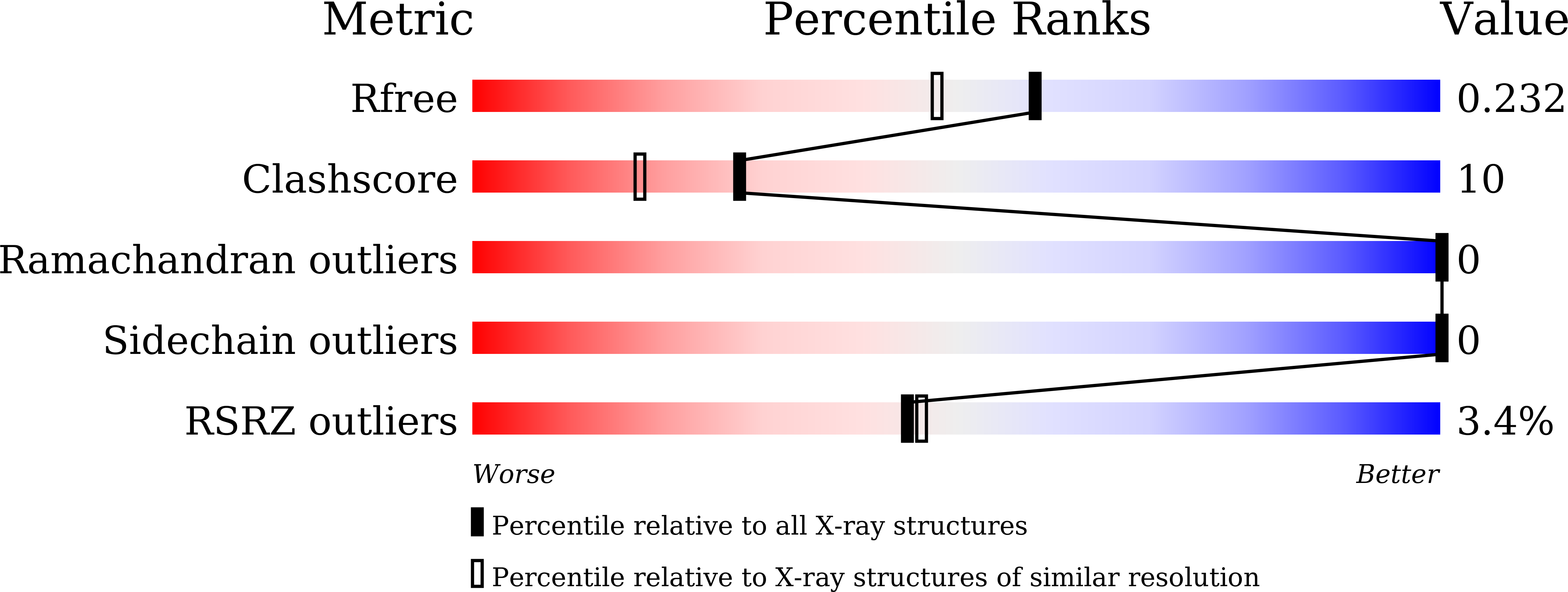
Experiments and Validation Details

X-ray source:
SSRF BEAMLINE BL17U1
Spacegroup:
P63
Expression system: Escherichia coli BL21