


Function and Biology Details
Biochemical function:
Biological process:
Cellular component:
Sequence domains:
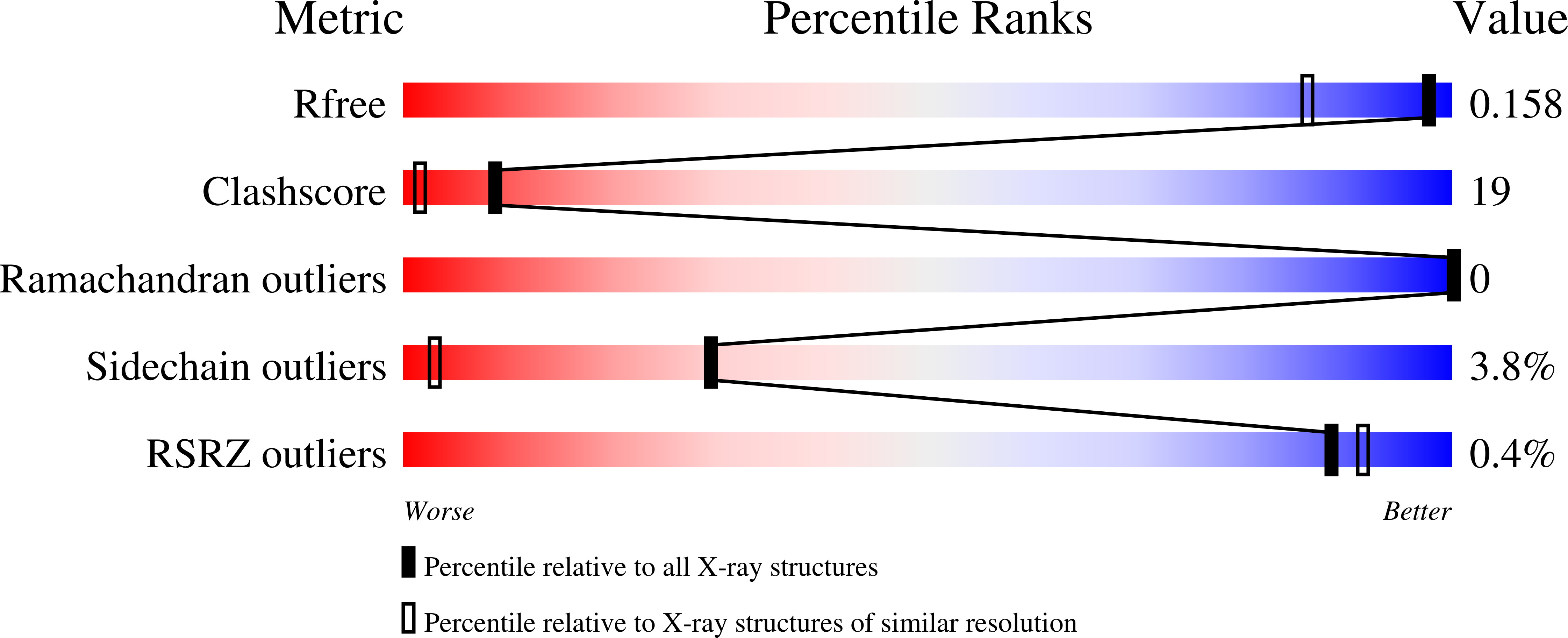
Structure analysis Details
Assembly composition:
homo trimer (preferred)
Assembly name:
Bacteriorhodopsin (preferred)
PDBe Complex ID:
PDB-CPX-136413 (preferred)
Entry contents:
1 distinct polypeptide molecule
Macromolecule:
Bacteriorhodopsin
Molecule details ›
Chain: A
Length: 229 amino acids
Theoretical weight: 25.06 KDa
Source organism: Halobacterium salinarum NRC-1
UniProt:
Sequence domains: Bacteriorhodopsin-like protein
Structure domains: Rhodopsin 7-helix transmembrane proteins
Length: 229 amino acids
Theoretical weight: 25.06 KDa
Source organism: Halobacterium salinarum NRC-1
UniProt:
- Canonical:
 P02945 (Residues: 18-246; Coverage: 87%)
Sequence domains: Bacteriorhodopsin-like protein
Structure domains: Rhodopsin 7-helix transmembrane proteins


