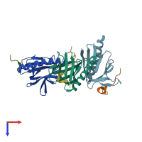
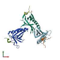
5zz9

X-ray diffraction
2.3Å resolution

Crystal structure of Homer2 EVH1/Drebrin PPXXF complex

Released:
Source organisms:
Primary publication:
Homer Tetramer Promotes Actin Bundling Activity of Drebrin.
Structure 27 27-38.e4 (2019)
PMID: 30503778

Function and Biology Details

Structure analysis Details

Assembly composition:
hetero dimer (preferred)
PDBe Complex ID:
PDB-CPX-172536 (preferred)
Entry contents:
2 distinct polypeptide molecules
Macromolecules (2 distinct):
Homer protein homolog 2 Chains: A, B, C
Molecule details ›
Chains: A, B, C
Length: 123 amino acids
Theoretical weight: 13.55 KDa
Source organism: Mus musculus
Expression system: Escherichia coli
UniProt:
  • Canonical: Q9QWW1 (Residues: 1-115; Coverage: 33%)
Gene names: Homer2, Vesl2
Sequence domains: WH1 domain
Structure domains: Pleckstrin-homology domain (PH domain)/Phosphotyrosine-binding domain (PTB)
Drebrin Chains: D, E, F
Molecule details ›
Chains: D, E, F
Length: 28 amino acids
Theoretical weight: 3.09 KDa
Source organism: Homo sapiens
Expression system: Escherichia coli
UniProt:
  • Canonical: Q16643 (Residues: 530-551; Coverage: 3%)
Gene names: D0S117E, DBN1

Ligands and Environments

No bound ligands
No modified residues

Experiments and Validation Details

Entry percentile scores
X-ray source: SSRF BEAMLINE BL19U1
Spacegroup: P1
Unit cell:
a: 45.337Å b: 47.808Å c: 50.415Å
α: 70.44° β: 83.56° γ: 90.39°
R-values:
R R work R free
0.193 0.192 0.228
Expression system: Escherichia coli