

-3,4-dihydro-2H,6H-pyrano[3,2-b]xanthen-6-one</span>;</li> <li class='image_legend_li'>1 copy of <span class='highlight'>ZINC ION</span>;</li> <li class='image_legend_li'>1 copy of <span class='highlight'>SULFATE ION</span>;</li> <li class='image_legend_li'>1 copy of <span class='highlight'>water</span>.</li></ul>")
-3,4-dihydro-2H,6H-pyrano[3,2-b]xanthen-6-one</span>;</li> <li class='image_legend_li'>1 copy of <span class='highlight'>ZINC ION</span>;</li> <li class='image_legend_li'>1 copy of <span class='highlight'>SULFATE ION</span>;</li> <li class='image_legend_li'>1 copy of <span class='highlight'>water</span>.</li></ul>")
-3,4-dihydro-2H,6H-pyrano[3,2-b]xanthen-6-one</span>;</li> <li class='image_legend_li'>1 copy of <span class='highlight'>ZINC ION</span>;</li> <li class='image_legend_li'>1 copy of <span class='highlight'>SULFATE ION</span>;</li> <li class='image_legend_li'>1 copy of <span class='highlight'>water</span>.</li></ul>")
Function and Biology Details
Reaction catalysed:
2-hydroxy-dATP + H(2)O = 2-hydroxy-dAMP + diphosphate
Biochemical function:
Biological process:
Cellular component:
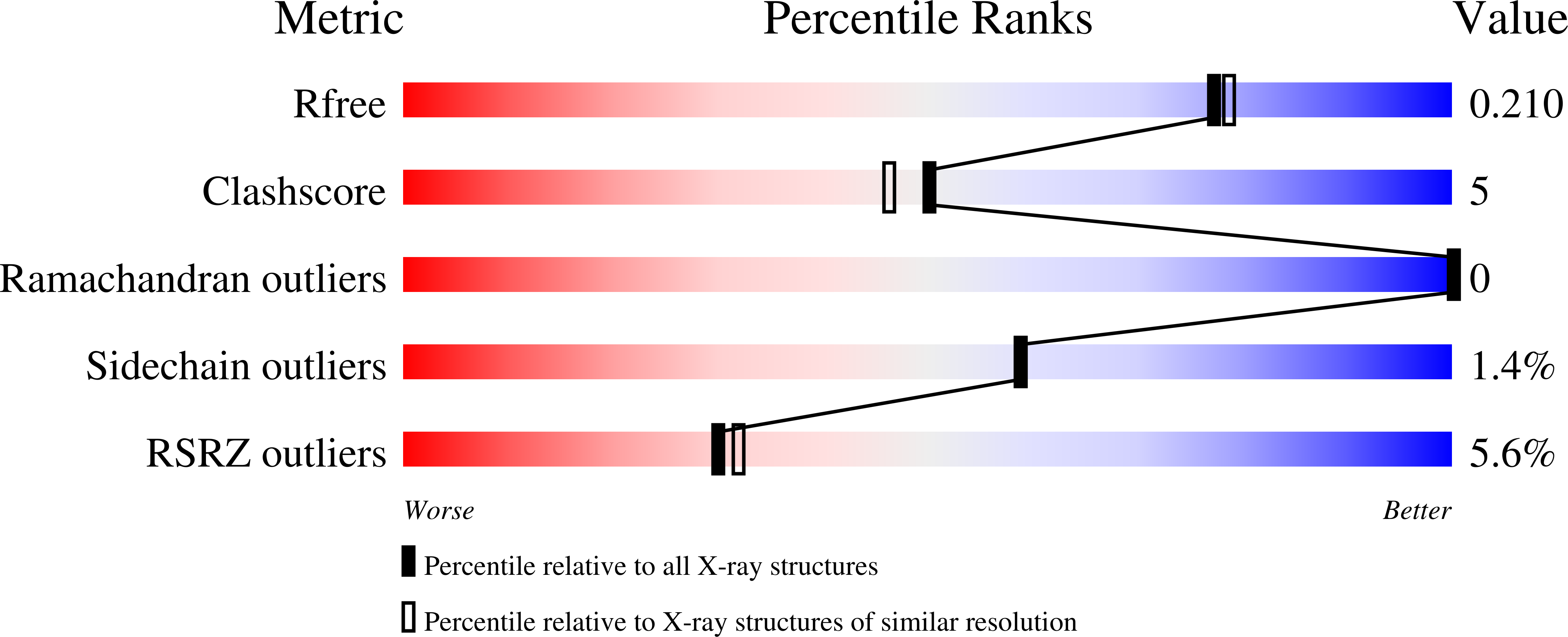
Structure analysis Details
Assemblies composition:
Assembly name:
PDBe Complex ID:
PDB-CPX-153202 (preferred)
Entry contents:
1 distinct polypeptide molecule
Macromolecule:
Oxidized purine nucleoside triphosphate hydrolase
Molecule details ›
Chain: A
Length: 163 amino acids
Theoretical weight: 18.99 KDa
Source organism: Homo sapiens
Expression system: Escherichia coli
UniProt:
Sequence domains: NUDIX domain
Length: 163 amino acids
Theoretical weight: 18.99 KDa
Source organism: Homo sapiens
Expression system: Escherichia coli
UniProt:
- Canonical:
 P36639 (Residues: 3-156; Coverage: 99%)
Sequence domains: NUDIX domain


