


Function and Biology Details
Reaction catalysed:
ATP + a [protein]-L-tyrosine = ADP + a [protein]-L-tyrosine phosphate
Biochemical function:
Biological process:
Cellular component:
- not assigned
Structure analysis Details
Assembly composition:
homo dimer (preferred)
Assembly name:
Inactive tyrosine-protein kinase PEAK1 (preferred)
PDBe Complex ID:
PDB-CPX-190658 (preferred)
Entry contents:
1 distinct polypeptide molecule
Macromolecule:
Inactive tyrosine-protein kinase PEAK1
Molecule details ›
Chain: A
Length: 438 amino acids
Theoretical weight: 50.15 KDa
Source organism: Homo sapiens
Expression system: Escherichia coli BL21(DE3)
UniProt:
Sequence domains: Protein kinase domain
Length: 438 amino acids
Theoretical weight: 50.15 KDa
Source organism: Homo sapiens
Expression system: Escherichia coli BL21(DE3)
UniProt:
- Canonical:
 Q9H792 (Residues: 1272-1743; Coverage: 25%)
Sequence domains: Protein kinase domain
Ligands and Environments
No bound ligands
No modified residues
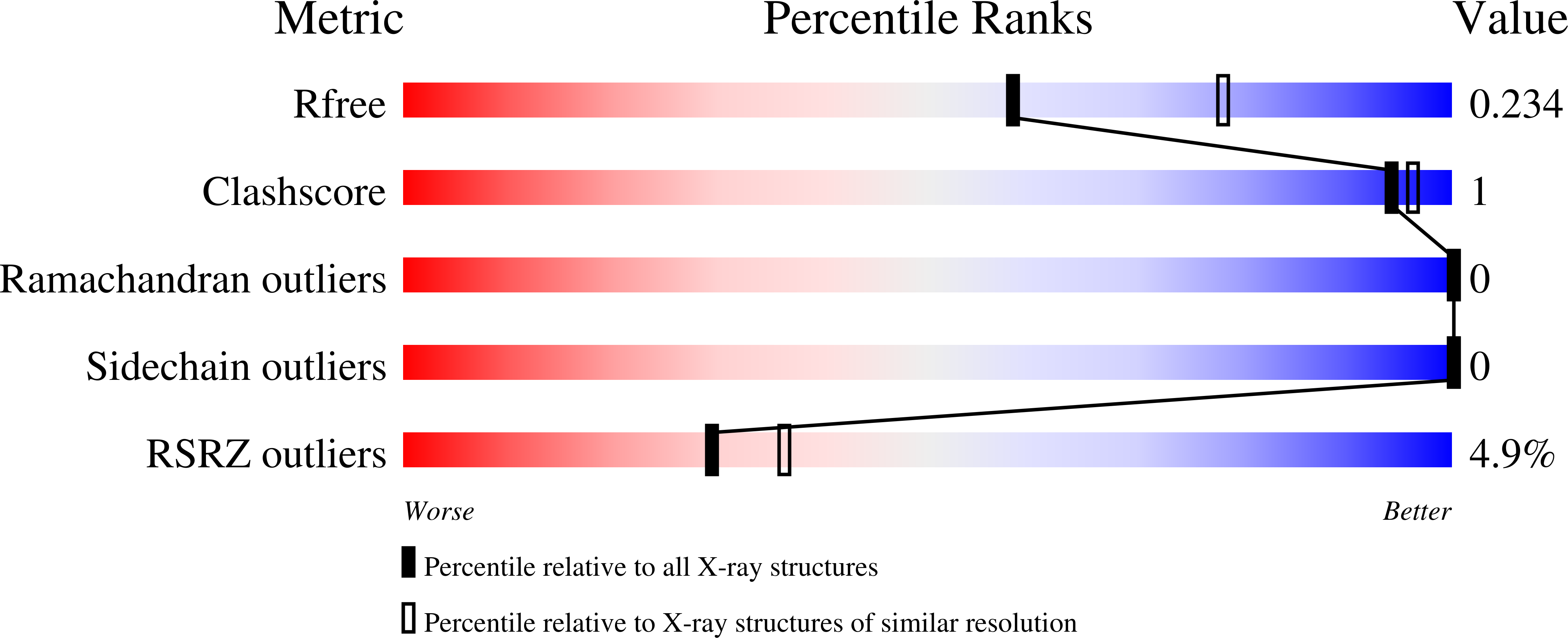
Experiments and Validation Details

X-ray source:
APS BEAMLINE 24-ID-E
Expression system: Escherichia coli BL21(DE3)