6bja

X-ray diffraction
1.6Å resolution

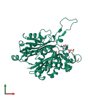
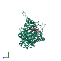
Crystal structure of Acat5 thiolase from Ascaris suum in complex with coenzyme A

Released:
Source organism: Ascaris suum
Primary publication:
Structural and Biochemical Studies of Substrate Selectivity in Ascaris suum Thiolases.
Biochemistry 57 3155-3166 (2018)
PMID: 29381332

Function and Biology Details

Structure analysis Details

Assembly composition:
homo tetramer (preferred)
PDBe Complex ID:
PDB-CPX-123297 (preferred)
Entry contents:
1 distinct polypeptide molecule
Macromolecule:
Acetyl-CoA acetyltransferase A Chain: A
Molecule details ›
Chain: A
Length: 397 amino acids
Theoretical weight: 41.99 KDa
Source organism: Ascaris suum
Expression system: Escherichia coli BL21(DE3)
UniProt:
  • Canonical: F1L3N8 (Residues: 28-421; Coverage: 94%)
Sequence domains:
Structure domains: Peroxisomal Thiolase; Chain A, domain 1

Ligands and Environments

2 bound ligands:
1 modified residue:

Experiments and Validation Details

Entry percentile scores
X-ray source: ALS BEAMLINE 8.3.1
Spacegroup: I222
Unit cell:
a: 71.662Å b: 111.78Å c: 112.756Å
α: 90° β: 90° γ: 90°
R-values:
R R work R free
0.147 0.146 0.173
Expression system: Escherichia coli BL21(DE3)