


Function and Biology Details
Reaction catalysed:
ATP + D-ribose = ADP + D-ribose 5-phosphate
Biochemical function:
Biological process:
Cellular component:
- not assigned
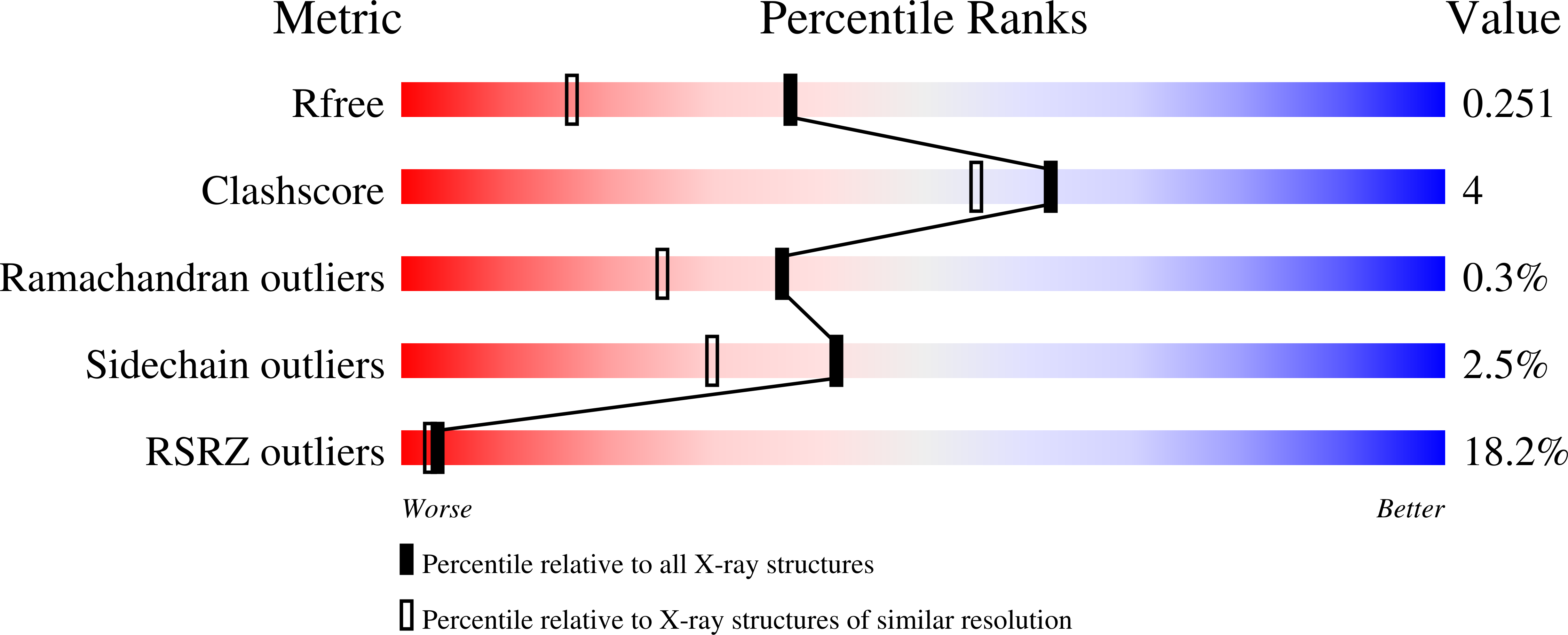
Structure analysis Details
Assembly composition:
homo dimer (preferred)
Assembly name:
Ribokinase (preferred)
PDBe Complex ID:
PDB-CPX-106545 (preferred)
Entry contents:
1 distinct polypeptide molecule
Macromolecule:
Ribokinase
Molecule details ›
Chains: A, B
Length: 313 amino acids
Theoretical weight: 32.93 KDa
Source organism: Arabidopsis thaliana
Expression system: Escherichia coli BL21(DE3)
UniProt:
Sequence domains: pfkB family carbohydrate kinase
Structure domains: UDP-N-acetylmuramoyl-L-alanine:D-glutamate ligase
Length: 313 amino acids
Theoretical weight: 32.93 KDa
Source organism: Arabidopsis thaliana
Expression system: Escherichia coli BL21(DE3)
UniProt:
- Canonical:
 A1A6H3 (Residues: 68-379; Coverage: 82%)
Sequence domains: pfkB family carbohydrate kinase
Structure domains: UDP-N-acetylmuramoyl-L-alanine:D-glutamate ligase


