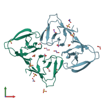
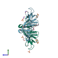
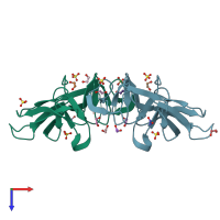
6ilu

X-ray diffraction
1.6Å resolution

Endolysin LysPBC5 CBD

Released:
Source organism: Bacillus phage PBC5
Primary publication:
Structural Basis for Cell-Wall Recognition by Bacteriophage PBC5 Endolysin.
Structure 27 1355-1365.e4 (2019)
PMID: 31353242

Function and Biology Details

Reaction catalysed:
Hydrolysis of (1->4)-beta-linkages between N-acetylmuramic acid and N-acetyl-D-glucosamine residues in a peptidoglycan and between N-acetyl-D-glucosamine residues in chitodextrins
Biochemical function:
  • not assigned
Biological process:
  • not assigned
Cellular component:
  • not assigned

Structure analysis Details

Assembly composition:
monomeric (preferred)
Assembly name:
PDBe Complex ID:
PDB-CPX-103925 (preferred)
Entry contents:
1 distinct polypeptide molecule
Macromolecule:
lysozyme Chains: A, B
Molecule details ›
Chains: A, B
Length: 140 amino acids
Theoretical weight: 15.52 KDa
Source organism: Bacillus phage PBC5
Expression system: Escherichia coli BL21(DE3)
UniProt:
  • Canonical: A0A218KCJ1 (Residues: 200-339; Coverage: 41%)
Gene name: PBC5_032

Ligands and Environments

2 bound ligands:
No modified residues

Experiments and Validation Details

Entry percentile scores
X-ray source: PAL/PLS BEAMLINE 5C (4A)
Spacegroup: I222
Unit cell:
a: 81.823Å b: 107.514Å c: 117.524Å
α: 90° β: 90° γ: 90°
R-values:
R R work R free
0.168 0.167 0.196
Expression system: Escherichia coli BL21(DE3)