


Function and Biology Details
Biochemical function:
Biological process:
Cellular component:
Structure analysis Details
Assembly composition:
homo dimer (preferred)
Assembly name:
Uridine phosphorylase (preferred)
PDBe Complex ID:
PDB-CPX-166965 (preferred)
Entry contents:
1 distinct polypeptide molecule
Macromolecule:
Ligands and Environments
No bound ligands
No modified residues
Experiments and Validation Details
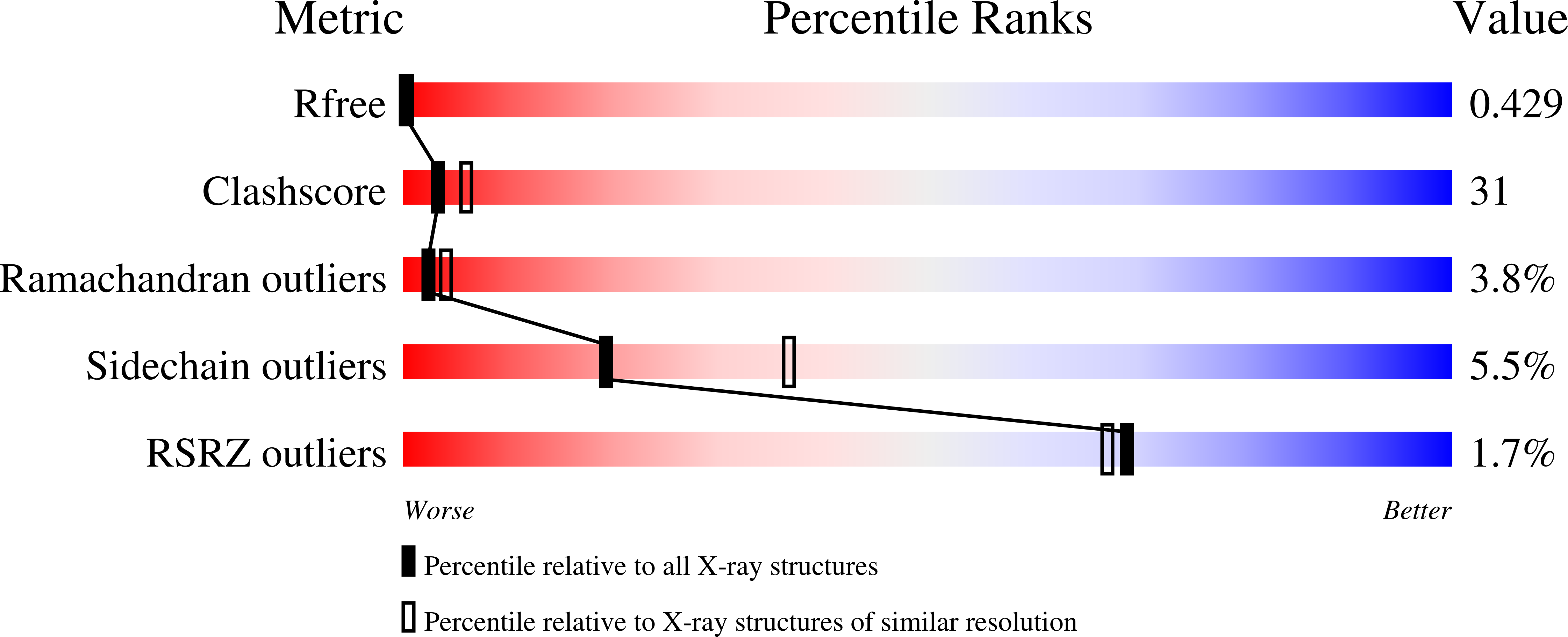
wwPDB Validation report is not available for this entry.
X-ray source:
SSRF BEAMLINE BL19U1
Spacegroup:
C2
Expression system: Escherichia coli
