


Function and Biology Details
Reaction catalysed:
[D-ribulose 5-phosphate + ATP = D-ribulose 1,5-bisphosphate + ADP + H(+)., D-ribulose 5-phosphate + ATP = D-ribulose 1,5-bisphosphate + ADP + H(+).]
Biochemical function:
Biological process:
Cellular component:
- not assigned
Structure analysis Details
Assembly composition:
homo dimer (preferred)
Assembly name:
Phosphoribulokinase, chloroplastic (preferred)
PDBe Complex ID:
PDB-CPX-150561 (preferred)
Entry contents:
1 distinct polypeptide molecule
Macromolecule:
Phosphoribulokinase, chloroplastic
Molecule details ›
Chains: A, B
Length: 359 amino acids
Theoretical weight: 40.5 KDa
Source organism: Arabidopsis thaliana
Expression system: Escherichia coli
UniProt:
Sequence domains: Phosphoribulokinase / Uridine kinase family
Length: 359 amino acids
Theoretical weight: 40.5 KDa
Source organism: Arabidopsis thaliana
Expression system: Escherichia coli
UniProt:
- Canonical:
 P25697 (Residues: 47-395; Coverage: 88%)
Sequence domains: Phosphoribulokinase / Uridine kinase family
Ligands and Environments
No bound ligands
No modified residues
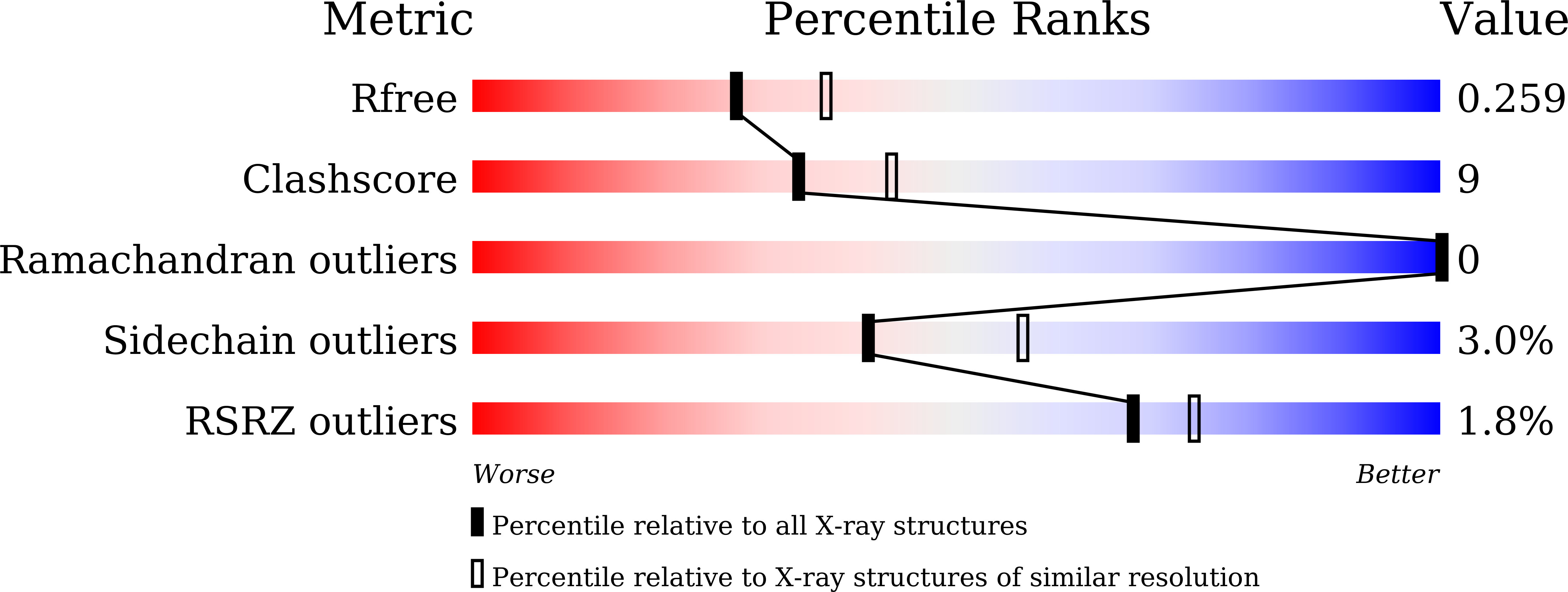
Experiments and Validation Details
wwPDB Validation report is not available for this entry.
X-ray source:
SSRF BEAMLINE BL17U
Spacegroup:
C2
Expression system: Escherichia coli
