6m3d

X-ray diffraction
1.6Å resolution

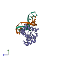
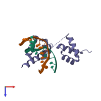
X-ray crystal structure of tandemly connected engrailed homeodomains (EHD) with R53A mutations and DNA complex

Released:
Source organism: Drosophila melanogaster
Primary publication:
Structural basis for designing an array of engrailed homeodomains.
Acta Crystallogr D Struct Biol 76 824-833 (2020)
PMID: 32876058

Function and Biology Details

Structure analysis Details

Assembly composition:
hetero trimer (preferred)
PDBe Complex ID:
PDB-CPX-120258 (preferred)
Entry contents:
1 distinct polypeptide molecule
2 distinct DNA molecules
Macromolecules (3 distinct):
Segmentation polarity homeobox protein engrailed Chain: C
Molecule details ›
Chain: C
Length: 148 amino acids
Theoretical weight: 17.34 KDa
Source organism: Drosophila melanogaster
Expression system: Escherichia coli BL21
UniProt:
  • Canonical: P02836 (Residues: 453-512; Coverage: 11%)
Gene names: CG9015, en
Sequence domains: Homeodomain
DNA (5'-D(*TP*AP*AP*TP*CP*CP*TP*AP*AP*TP*CP*C)-3') Chain: A
Molecule details ›
Chain: A
Length: 12 nucleotides
Theoretical weight: 3.58 KDa
DNA (5'-D(*GP*GP*AP*TP*TP*AP*GP*GP*AP*TP*TP*A)-3') Chain: B
Molecule details ›
Chain: B
Length: 12 nucleotides
Theoretical weight: 3.74 KDa

Ligands and Environments

1 bound ligand:
No modified residues

Experiments and Validation Details

Entry percentile scores
X-ray source: PHOTON FACTORY BEAMLINE BL-17A
Spacegroup: P21
Unit cell:
a: 39.798Å b: 61.437Å c: 39.976Å
α: 90° β: 106.447° γ: 90°
R-values:
R R work R free
0.191 0.188 0.236
Expression system: Escherichia coli BL21