6o8e

X-ray diffraction
2.61Å resolution

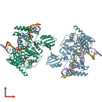
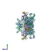
Crystal structure of UvrB bound to duplex DNA with ADP

Released:
Source organism: [Bacillus] caldotenax
Primary publication:
Mechanism of DNA Lesion Homing and Recognition by the Uvr Nucleotide Excision Repair System.
Research (Wash D C) 2019 5641746 (2019)
PMID: 31549070

Function and Biology Details

Structure analysis Details

Assembly composition:
hetero trimer (preferred)
PDBe Complex ID:
PDB-CPX-120368 (preferred)
Entry contents:
1 distinct polypeptide molecule
2 distinct DNA molecules
Macromolecules (3 distinct):
UvrABC system protein B Chains: A, B
Molecule details ›
Chains: A, B
Length: 593 amino acids
Theoretical weight: 68.14 KDa
Source organism: [Bacillus] caldotenax
Expression system: Escherichia coli
UniProt:
  • Canonical: P56981 (Residues: 2-190, 191-593; Coverage: 90%)
Gene name: uvrB
Sequence domains:
DNA (5'-D(*GP*CP*TP*CP*TP*AP*GP*AP*TP*TP*TP*TP*CP*AP*TP*AP*CP*GP*GP*C)-3') Chains: C, E
Molecule details ›
Chains: C, E
Length: 20 nucleotides
Theoretical weight: 6.1 KDa
DNA (5'-D(*GP*CP*CP*GP*TP*AP*TP*GP*CP*CP*AP*AP*TP*CP*TP*AP*GP*AP*GP*C)-3') Chains: D, F
Molecule details ›
Chains: D, F
Length: 20 nucleotides
Theoretical weight: 6.12 KDa

Ligands and Environments

No modified residues

Experiments and Validation Details

Entry percentile scores
X-ray source: APS BEAMLINE 24-ID-E
Unit cell:
a: 69.269Å b: 115.719Å c: 266.795Å
α: 90° β: 90° γ: 90°
R-values:
R R work R free
0.218 0.215 0.27
Expression system: Escherichia coli