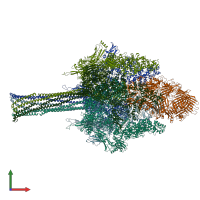
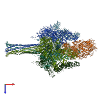
6suf

Electron Microscopy
3.4Å resolution

Structure of Photorhabdus luminescens Tc holotoxin pore

Released:
Source organism: Photorhabdus luminescens
Primary publication:
Structure of a Tc holotoxin pore provides insights into the translocation mechanism.
Proc Natl Acad Sci U S A 116 23083-23090 (2019)
PMID: 31666324
Related structures: EMD-10313

Function and Biology Details

Structure analysis Details

Assembly composition:
hetero hexamer (preferred)
Assembly name:
PDBe Complex ID:
PDB-CPX-184505 (preferred)
Entry contents:
2 distinct polypeptide molecules
Macromolecules (2 distinct):
TcdA1 Chains: A, B, C, D, E
Molecule details ›
Chains: A, B, C, D, E
Length: 2516 amino acids
Theoretical weight: 283.23 KDa
Source organism: Photorhabdus luminescens
Expression system: Escherichia coli
UniProt:
  • Canonical: Q9RN43 (Residues: 1-2516; Coverage: 100%)
Gene names: tcdA, tcdA1
Sequence domains:
TccC3; TcdB2 Chain: F
Molecule details ›
Chain: F
Length: 2439 amino acids
Theoretical weight: 274.19 KDa
Source organism: Photorhabdus luminescens
Expression system: Escherichia coli
UniProt:
  • Canonical: Q8GF99 (Residues: 1-1474; Coverage: 100%)
  • Canonical: Q8GF97 (Residues: 1-960; Coverage: 100%)
Gene names: TccC3, tcdB2
Sequence domains:

Ligands and Environments

No bound ligands
No modified residues

Experiments and Validation Details

Entry percentile scores
Resolution: 3.4Å
Relevant EMDB volumes: EMD-10313
Expression system: Escherichia coli