We've built a new version of this page with an interactive 3D viewer. Try it out in beta and help us improve:

6svj

X-ray diffraction
1.16Å resolution

Terahertz irradiated structure of bovine trypsin (odd frames of crystal x42)

Released:
Model geometry
Fit model/data

Function and Biology Details

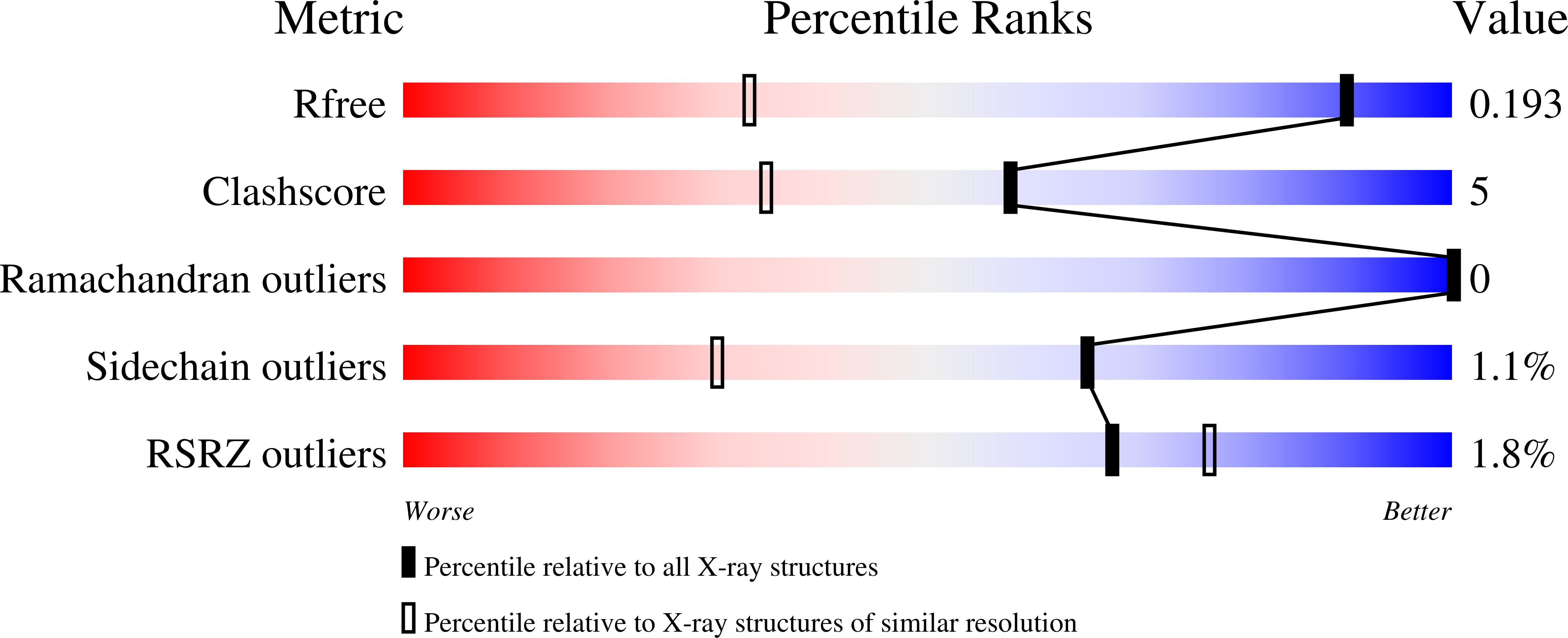
Structure analysis Details

Assembly composition:
monomeric (preferred)
Assembly name:
PDBe Complex ID:
PDB-CPX-133319 (preferred)
Entry contents:
1 distinct polypeptide molecule
Macromolecule:
Serine protease 1 Chain: A
Molecule details ›
Chain: A
Length: 223 amino acids
Theoretical weight: 23.32 KDa
Source organism: Bos taurus
UniProt:
  • Canonical: P00760 (Residues: 24-246; Coverage: 97%)
Gene names: PRSS1, TRP1, TRY1, TRYP1
Sequence domains: Trypsin

Ligands and Environments

3 bound ligands:
No modified residues

Experiments and Validation Details

wwPDB Validation report is not available for this entry.
X-ray source: PETRA III, EMBL c/o DESY BEAMLINE P14 (MX2)
Spacegroup: P212121
Unit cell:
a: 53.91Å b: 57.15Å c: 65.7Å
α: 90° β: 90° γ: 90°
R-values:
R R work R free
0.15 0.15 0.192