


Function and Biology Details
Reaction catalysed:
Endohydrolysis of (1->4)-beta-D-glucosidic linkages in cellulose, lichenin and cereal beta-D-glucans
Biochemical function:
Biological process:
Cellular component:
- not assigned
Structure analysis Details
Assembly composition:
monomeric (preferred)
Assembly name:
Endoglucanase (preferred)
PDBe Complex ID:
PDB-CPX-145314 (preferred)
Entry contents:
1 distinct polypeptide molecule
Macromolecule:
Endoglucanase
Molecule details ›
Chain: A
Length: 149 amino acids
Theoretical weight: 16.72 KDa
Source organism: Bacillus subtilis subsp. subtilis str. 168
Expression system: Escherichia coli
UniProt:
Sequence domains: Cellulose binding domain
Length: 149 amino acids
Theoretical weight: 16.72 KDa
Source organism: Bacillus subtilis subsp. subtilis str. 168
Expression system: Escherichia coli
UniProt:
- Canonical:
 P10475 (Residues: 354-499; Coverage: 31%)
Sequence domains: Cellulose binding domain
Ligands and Environments
No bound ligands
No modified residues
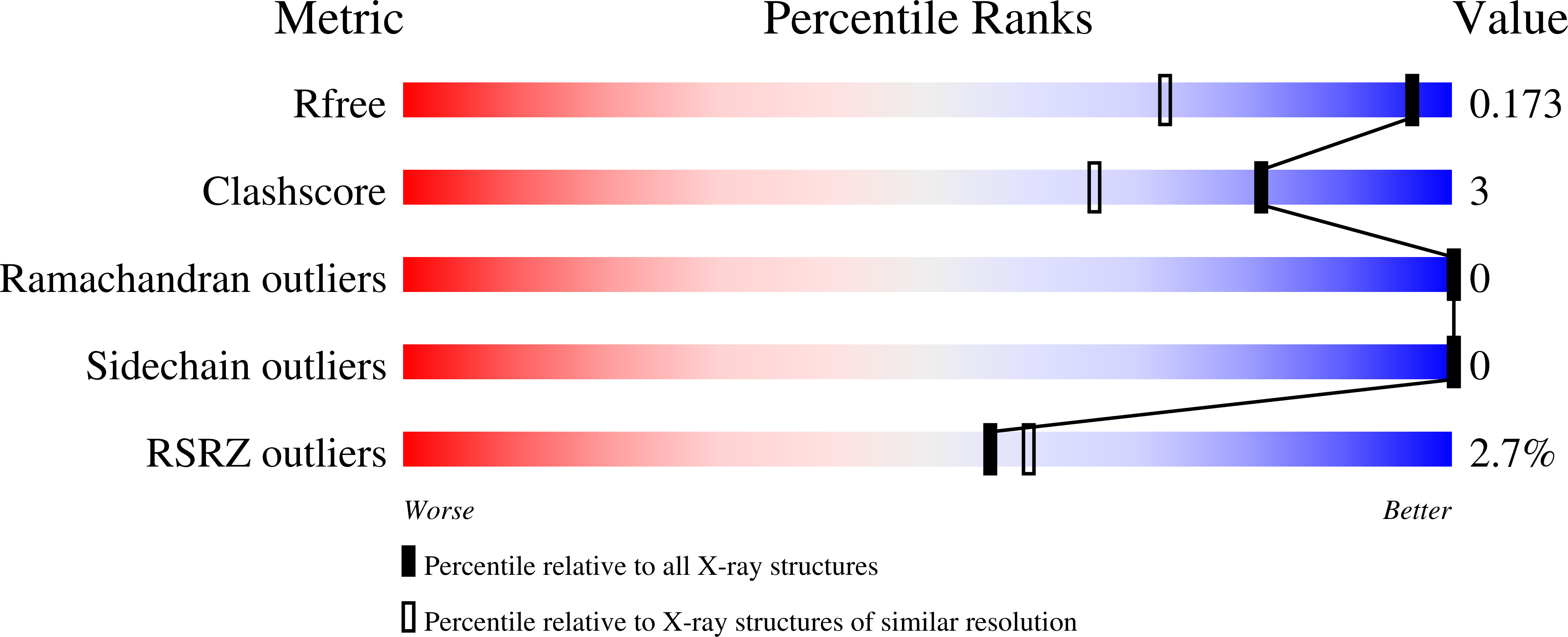
Experiments and Validation Details
wwPDB Validation report is not available for this entry.
X-ray source:
LNLS BEAMLINE W01B-MX2
Spacegroup:
C2221
Expression system: Escherichia coli
