

-2-hydroxybutanedioic acid</span>;</li> <li class='image_legend_li'>2 copies of <span class='highlight'>GLYCEROL</span>;</li> <li class='image_legend_li'>1 copy of <span class='highlight'>water</span>.</li></ul>")
-2-hydroxybutanedioic acid</span>;</li> <li class='image_legend_li'>2 copies of <span class='highlight'>GLYCEROL</span>;</li> <li class='image_legend_li'>1 copy of <span class='highlight'>water</span>.</li></ul>")
-2-hydroxybutanedioic acid</span>;</li> <li class='image_legend_li'>2 copies of <span class='highlight'>GLYCEROL</span>;</li> <li class='image_legend_li'>1 copy of <span class='highlight'>water</span>.</li></ul>")

Function and Biology Details
Reaction catalysed:
(S)-malate = fumarate + H(2)O
Biochemical function:
Biological process:
Cellular component:
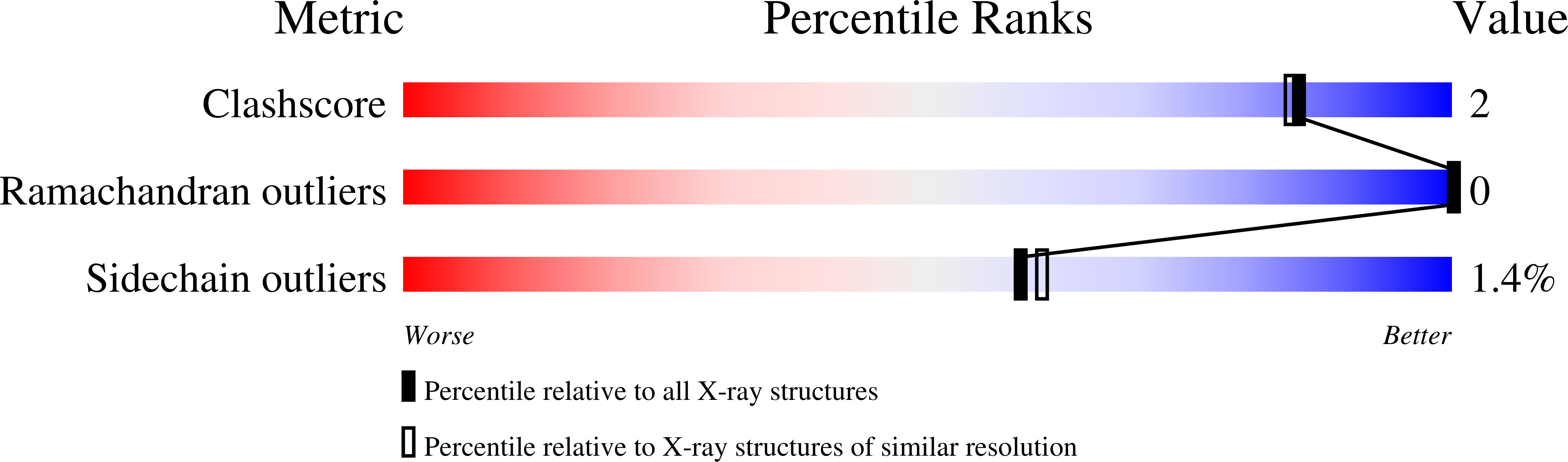
Structure analysis Details
Assembly composition:
homo dimer (preferred)
Assembly name:
Fumarate hydratase 2 (preferred)
PDBe Complex ID:
PDB-CPX-123143 (preferred)
Entry contents:
1 distinct polypeptide molecule
Macromolecule:
Fumarate hydratase 2
Molecule details ›
Chains: A, B
Length: 604 amino acids
Theoretical weight: 66.48 KDa
Source organism: Leishmania major strain Friedlin
Expression system: Escherichia coli
UniProt:
Sequence domains:
Length: 604 amino acids
Theoretical weight: 66.48 KDa
Source organism: Leishmania major strain Friedlin
Expression system: Escherichia coli
UniProt:
- Canonical:
 E9AE57 (Residues: 1-568; Coverage: 100%)
Sequence domains:


