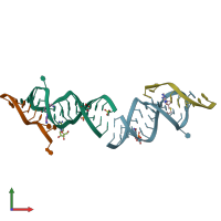
6yml

X-ray diffraction
2.17Å resolution

Crystal structure of the SAM-SAH riboswitch with decarboxylated SAH

Released:
Source organism: Roseobacter sp.
Primary publication:
Crystal structure and ligand-induced folding of the SAM/SAH riboswitch.
Nucleic Acids Res 48 7545-7556 (2020)
PMID: 32520325

Function and Biology Details

Biochemical function:
  • not assigned
Biological process:
  • not assigned
Cellular component:
  • not assigned

Structure analysis Details

Assembly composition:
hetero dimer (preferred)
Assembly name:
PDBe Complex ID:
PDB-CPX-197188 (preferred)
Entry contents:
2 distinct RNA molecules
Macromolecules (2 distinct):
Chains: A,C Chains: A, C
Molecule details ›
Chains: A, C
Length: 26 nucleotides
Theoretical weight: 8.39 KDa
Source organism: Roseobacter sp.
Expression system: Not provided
Chains: B,D Chains: B, D
Molecule details ›
Chains: B, D
Length: 9 nucleotides
Theoretical weight: 2.9 KDa
Source organism: Roseobacter sp.
Expression system: Not provided

Ligands and Environments

3 bound ligands:
1 modified residue:

Experiments and Validation Details

Entry percentile scores
X-ray source: DIAMOND BEAMLINE I24
Spacegroup: P312
Unit cell:
a: 86.218Å b: 86.218Å c: 75.395Å
α: 90° β: 90° γ: 120°
R-values:
R R work R free
0.2 0.198 0.233
Expression system: Not provided