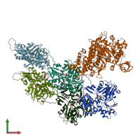
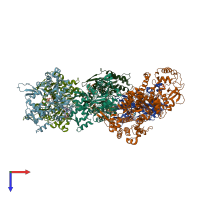
7aln

Electron Microscopy
3.77Å resolution

Cryo-EM structure of the divergent actomyosin complex from Plasmodium falciparum Myosin A in the Rigor state

Released:

Function and Biology Details

Structure analysis Details

Assembly composition:
hetero hexamer (preferred)
Assembly name:
PDBe Complex ID:
PDB-CPX-184680 (preferred)
Entry contents:
2 distinct polypeptide molecules
Macromolecules (2 distinct):
Actin-1 Chains: A, B, C, D, E
Molecule details ›
Chains: A, B, C, D, E
Length: 376 amino acids
Theoretical weight: 41.92 KDa
Source organism: Plasmodium falciparum 3D7
Expression system: Spodoptera frugiperda
UniProt:
  • Canonical: Q8I4X0 (Residues: 1-376; Coverage: 100%)
Gene names: ACT1, ACTI, PF3D7_1246200, PFL2215w
Sequence domains: Actin
Myosin-A Chain: F
Molecule details ›
Chain: F
Length: 818 amino acids
Theoretical weight: 92.49 KDa
Source organism: Plasmodium falciparum 3D7
Expression system: Spodoptera frugiperda
UniProt:
  • Canonical: Q8IDR3 (Residues: 1-818; Coverage: 100%)
Gene names: MyoA, PF13_0233, PF3D7_1342600
Sequence domains: Myosin head (motor domain)

Ligands and Environments

3 bound ligands:
1 modified residue:

Experiments and Validation Details

Entry percentile scores
Resolution: 3.77Å
Relevant EMDB volumes: EMD-11818
Expression system: Spodoptera frugiperda