


Function and Biology Details
Reaction catalysed:
GTP + H(2)O = GDP + phosphate
Biochemical function:
Biological process:
Cellular component:
Sequence domains:
- Translational (tr)-type GTP-binding domain
- Translation elongation factor EF1B, beta chain, archaeal
- Translation elongation factor EF1B, beta/delta subunit, guanine nucleotide exchange domain
- P-loop containing nucleoside triphosphate hydrolase
- Translation elongation factor EF1A, eukaryotic/archaeal
- Translation elongation factor eEF-1beta-like superfamily
- Translation elongation factor EF1B/small ribosomal subunit protein bS6
- Translation elongation factor EF1A/initiation factor IF2gamma, C-terminal
5 more domains
Structure analysis Details
Assembly composition:
hetero dimer (preferred)
Assembly name:
PDBe Complex ID:
PDB-CPX-129814 (preferred)
Entry contents:
2 distinct polypeptide molecules
Macromolecules (2 distinct):
Elongation factor 1-alpha
Molecule details ›
Chains: A, B
Length: 434 amino acids
Theoretical weight: 48.43 KDa
Source organism: Pyrococcus horikoshii OT3
Expression system: Escherichia coli
UniProt:
Sequence domains:
Length: 434 amino acids
Theoretical weight: 48.43 KDa
Source organism: Pyrococcus horikoshii OT3
Expression system: Escherichia coli
UniProt:
- Canonical:
 O59153 (Residues: 1-428; Coverage: 100%)
Sequence domains:
Elongation factor 1-beta
Molecule details ›
Chains: C, D
Length: 91 amino acids
Theoretical weight: 10.36 KDa
Source organism: Pyrococcus horikoshii OT3
Expression system: Escherichia coli
UniProt:
Sequence domains: EF-1 guanine nucleotide exchange domain
Length: 91 amino acids
Theoretical weight: 10.36 KDa
Source organism: Pyrococcus horikoshii OT3
Expression system: Escherichia coli
UniProt:
- Canonical: P58748 (Residues: 1-91; Coverage: 100%)
Sequence domains: EF-1 guanine nucleotide exchange domain
Ligands and Environments
No bound ligands
No modified residues
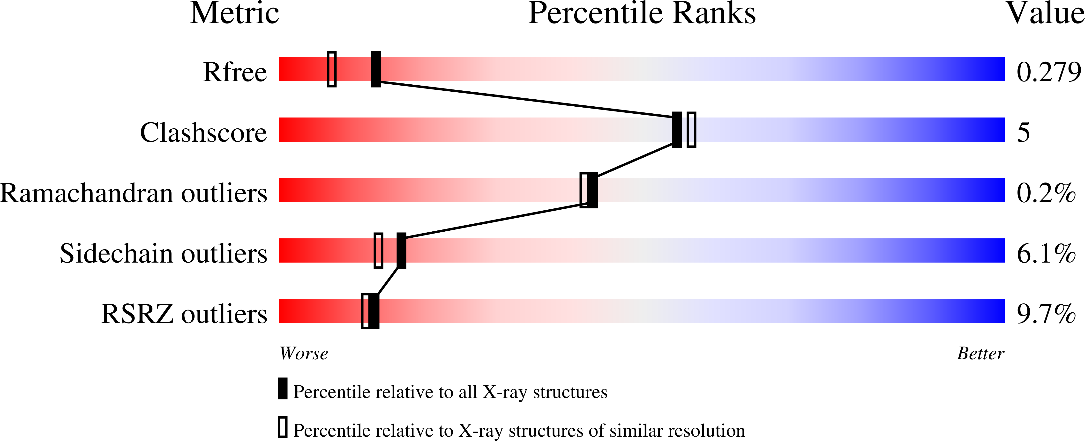
Experiments and Validation Details
wwPDB Validation report is not available for this entry.
X-ray source:
PHOTON FACTORY BEAMLINE AR-NW12A
Spacegroup:
P1
Expression system: Escherichia coli
