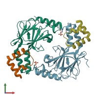
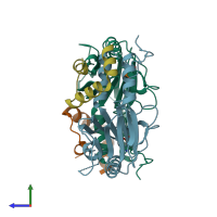
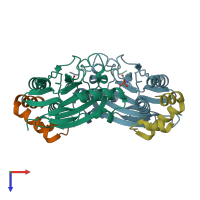
Function and Biology Details
Reaction catalysed:
Acetyl-CoA + [protein]-L-lysine = CoA + [protein]-N(6)-acetyl-L-lysine
Biochemical function:
Biological process:
Cellular component:
Sequence domains:
- Interferon regulatory factor-3
- Nucleolar pre-ribosomal-associated protein 1, N-terminal
- Nuclear receptor coactivator, CREB-bp-like, interlocking domain superfamily
- Nuclear receptor coactivator, CREB-bp-like, interlocking
- Nuclear receptor coactivator, interlocking
- SMAD/FHA domain superfamily
- SMAD-like domain superfamily
Structure analysis Details
Assembly composition:
hetero tetramer (preferred)
Assembly name:
PDBe Complex ID:
PDB-CPX-172057 (preferred)
Entry contents:
2 distinct polypeptide molecules
Macromolecules (2 distinct):


