7l6n

Electron Microscopy
7Å resolution

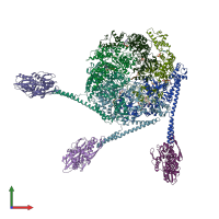
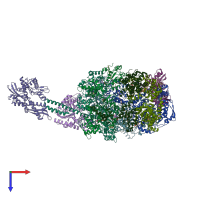
The Mycobacterium tuberculosis ClpB disaggregase hexamer structure with three locally refined ClpB middle domains and three DnaK nucleotide binding domains

Released:

Function and Biology Details

Structure analysis Details

Assembly composition:
hetero decamer (preferred)
PDBe Complex ID:
PDB-CPX-161800 (preferred)
Entry contents:
3 distinct polypeptide molecules
Macromolecules (3 distinct):
Chaperone protein ClpB Chains: A, B, C, D, E, F
Molecule details ›
Chains: A, B, C, D, E, F
Length: 848 amino acids
Theoretical weight: 92.69 KDa
Source organism: Mycobacterium tuberculosis
Expression system: Escherichia coli
UniProt:
  • Canonical: P9WPD1 (Residues: 1-848; Coverage: 100%)
Gene names: MTV036.19c, Rv0384c, clpB
Sequence domains:
Substrate Chain: N
Molecule details ›
Chain: N
Length: 33 amino acids
Theoretical weight: 2.83 KDa
Source organism: Mycobacterium tuberculosis
Expression system: Escherichia coli
Chaperone protein DnaK Chains: I, J, K
Molecule details ›
Chains: I, J, K
Length: 625 amino acids
Theoretical weight: 66.91 KDa
Source organism: Mycobacterium tuberculosis
Expression system: Escherichia coli
UniProt:
  • Canonical: P9WMJ9 (Residues: 1-625; Coverage: 100%)
Gene names: MTCY13E10.10, Rv0350, dnaK
Sequence domains: Hsp70 protein

Ligands and Environments

2 bound ligands:
No modified residues

Experiments and Validation Details

Entry percentile scores
Resolution:
Relevant EMDB volumes: EMD-23206
Expression system: Escherichia coli