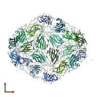
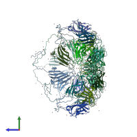
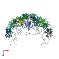
7m54

X-ray diffraction
3.8Å resolution

Crystallographic structure of a cubic crystal form of STMV grown from bromide

Released:
Primary publication:
Structures of additional crystal forms of Satellite tobacco mosaic virus grown from a variety of salts.
Acta Crystallogr F Struct Biol Commun 77 473-483 (2021)
PMID: 34866603

Function and Biology Details

Biochemical function:
Biological process:
  • not assigned
Cellular component:

Structure analysis Details

Assembly composition:
homo 60-mer (preferred)
Assembly name:
PDBe Complex ID:
PDB-CPX-148090 (preferred)
Entry contents:
1 distinct polypeptide molecule
Macromolecule:
Coat protein Chains: A, B, C, D, E, F, G, GG, H, HH, I, II, J, JJ, K, KK, L, M, N, O
Molecule details ›
Chains: A, B, C, D, E, F, G, GG, H, HH, I, II, J, JJ, K, KK, L, M, N, O
Length: 159 amino acids
Theoretical weight: 17.53 KDa
Source organism: Satellite tobacco mosaic virus
UniProt:
  • Canonical: P17574 (Residues: 1-159; Coverage: 100%)

Ligands and Environments

No modified residues

Experiments and Validation Details

Entry percentile scores
X-ray source: ALS BEAMLINE 8.3.1
Spacegroup: P23
Unit cell:
a: 234.65Å b: 234.65Å c: 234.65Å
α: 90° β: 90° γ: 90°
R-values:
R R work R free
0.219 0.213 0.285