7njz

X-ray diffraction
3.2Å resolution

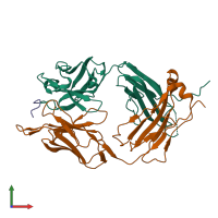
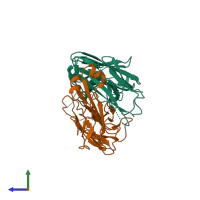
X-ray crystallography study of RoAb13 which binds to PIYDIN, a part of the CCR5 N terminal domain

Released:

Function and Biology Details

Structure analysis Details

Assembly composition:
hetero trimer (preferred)
PDBe Complex ID:
PDB-CPX-211299 (preferred)
Entry contents:
3 distinct polypeptide molecules
Macromolecules (3 distinct):
Antibody RoAb13 Heavy Chain Chain: H
Molecule details ›
Chain: H
Length: 224 amino acids
Theoretical weight: 24.06 KDa
Source organism: Mus musculus
Antibody RoAb13 Light Chain Chain: L
Molecule details ›
Chain: L
Length: 220 amino acids
Theoretical weight: 24.46 KDa
Source organism: Mus musculus
C-C chemokine receptor type 5 Chain: C
Molecule details ›
Chain: C
Length: 6 amino acids
Theoretical weight: 734 Da
Source organism: Homo sapiens
Expression system: Not provided
UniProt:
  • Canonical: P51681 (Residues: 8-13; Coverage: 2%)
Gene names: CCR5, CMKBR5

Ligands and Environments

No bound ligands
No modified residues

Experiments and Validation Details

Entry percentile scores
X-ray source: DIAMOND BEAMLINE I04
Spacegroup: P41212
Unit cell:
a: 76.44Å b: 76.44Å c: 268.639Å
α: 90° β: 90° γ: 90°
R-values:
R R work R free
0.27 0.269 0.292
Expression system: Not provided