7zfr

X-ray diffraction
2.9Å resolution

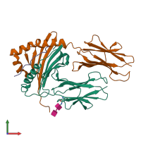
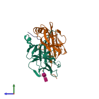
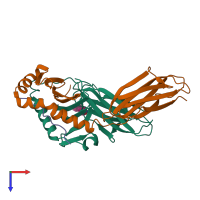
Crystal structure of HLA-DP (DPA1*02:01-DPB1*01:01) in complex with a peptide bound in the reverse direction

Released:

Function and Biology Details

Structure analysis Details

Assembly composition:
hetero trimer (preferred)
PDBe Complex ID:
PDB-CPX-199906 (preferred)
Entry contents:
3 distinct polypeptide molecules
Macromolecules (4 distinct):
MHC class II HLA-DP alpha chain (DPA1*02:01) Chain: A
Molecule details ›
Chain: A
Length: 268 amino acids
Theoretical weight: 29.81 KDa
Source organism: Homo sapiens
Expression system: Drosophila melanogaster
MHC class II HLA-DP beta chain (DPB1*01:01) Chain: B
Molecule details ›
Chain: B
Length: 262 amino acids
Theoretical weight: 29.73 KDa
Source organism: Homo sapiens
Expression system: Drosophila melanogaster
Synthetic peptide Chain: C
Molecule details ›
Chain: C
Length: 12 amino acids
Theoretical weight: 1.47 KDa
Source organism: Homo sapiens
Expression system: Not provided

Ligands and Environments

Carbohydrate polymer : NEW Components: NAG
No bound ligands
No modified residues

Experiments and Validation Details

Entry percentile scores
X-ray source: SLS BEAMLINE X06DA
Spacegroup: P6122
Unit cell:
a: 83.985Å b: 83.985Å c: 348.589Å
α: 90° β: 90° γ: 120°
R-values:
R R work R free
0.235 0.23 0.272
Expression systems:
  • Drosophila melanogaster
  • Not provided