


Function and Biology Details
Reaction catalysed:
dTDP-alpha-D-glucose = dTDP-4-dehydro-6-deoxy-alpha-D-glucose + H(2)O
Biochemical function:
Biological process:
Cellular component:
- not assigned
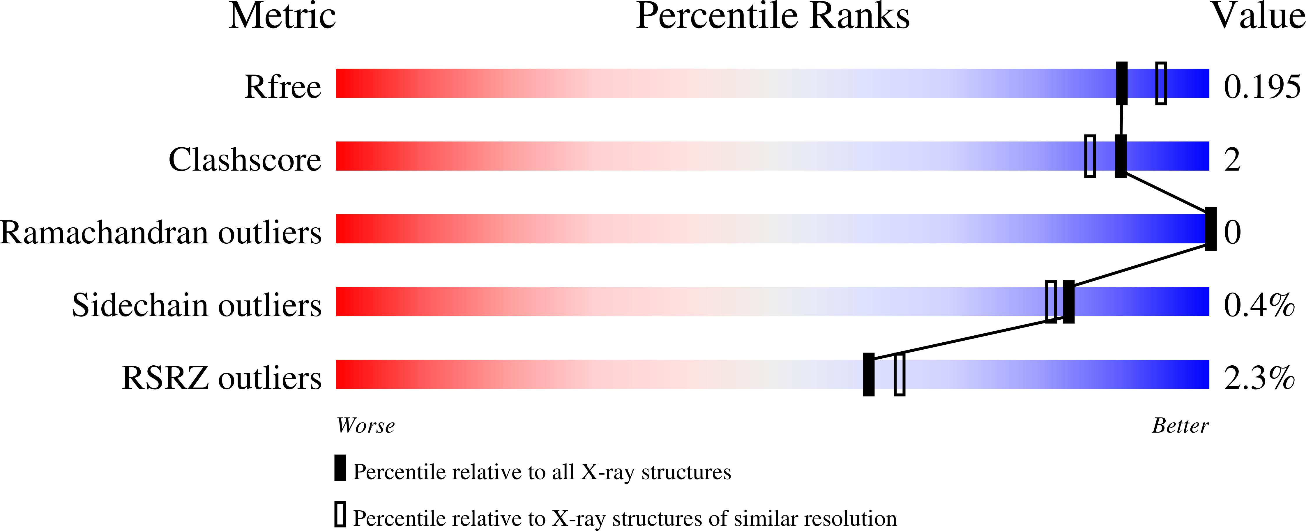
Structure analysis Details
Assembly composition:
homo dimer (preferred)
Assembly name:
dTDP-glucose 4,6-dehydratase (preferred)
PDBe Complex ID:
PDB-CPX-100520 (preferred)
Entry contents:
1 distinct polypeptide molecule
Macromolecule:
dTDP-glucose 4,6-dehydratase
Molecule details ›
Chains: A, B, C, D
Length: 367 amino acids
Theoretical weight: 42 KDa
Source organism: Elizabethkingia anophelis NUHP1
Expression system: Escherichia coli BL21(DE3)
UniProt:
Sequence domains: GDP-mannose 4,6 dehydratase
Length: 367 amino acids
Theoretical weight: 42 KDa
Source organism: Elizabethkingia anophelis NUHP1
Expression system: Escherichia coli BL21(DE3)
UniProt:
- Canonical:
 A0A077ELH2 (Residues: 1-359; Coverage: 100%)
Sequence domains: GDP-mannose 4,6 dehydratase


