

TRI(ETHYLOXY)OCTANE</span>;</li> <li class='image_legend_li'>3 copies of <span class='highlight'>LAURYL DIMETHYLAMINE-N-OXIDE</span>;</li> <li class='image_legend_li'>1 copy of <span class='highlight'>water</span>.</li></ul>")
TRI(ETHYLOXY)OCTANE</span>;</li> <li class='image_legend_li'>3 copies of <span class='highlight'>LAURYL DIMETHYLAMINE-N-OXIDE</span>;</li> <li class='image_legend_li'>1 copy of <span class='highlight'>water</span>.</li></ul>")
TRI(ETHYLOXY)OCTANE</span>;</li> <li class='image_legend_li'>3 copies of <span class='highlight'>LAURYL DIMETHYLAMINE-N-OXIDE</span>;</li> <li class='image_legend_li'>1 copy of <span class='highlight'>water</span>.</li></ul>")

Function and Biology Details
Biochemical function:
Biological process:
Cellular component:
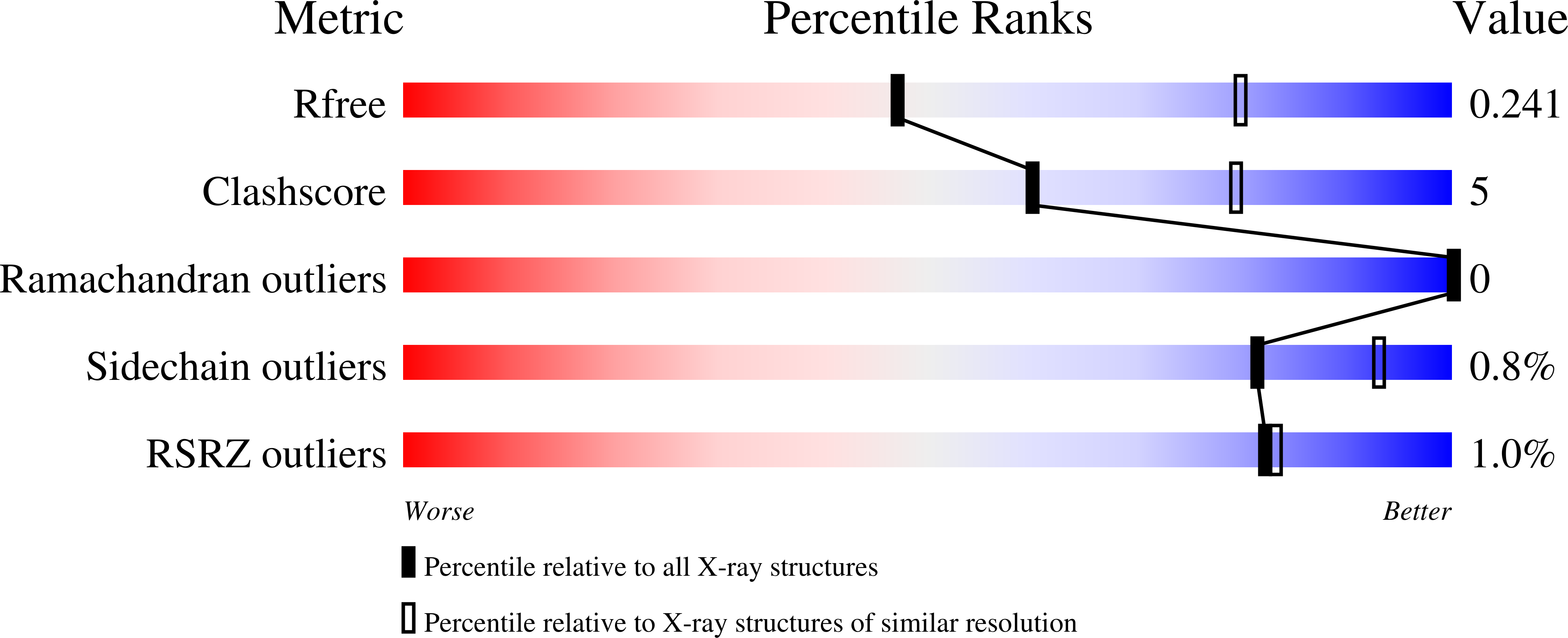
Structure analysis Details
Assembly composition:
homo trimer (preferred)
Assembly name:
OmpK36 (preferred)
PDBe Complex ID:
PDB-CPX-112798 (preferred)
Entry contents:
1 distinct polypeptide molecule
Macromolecule:
OmpK36
Molecule details ›
Chains: C, E, F
Length: 347 amino acids
Theoretical weight: 38.23 KDa
Source organism: Klebsiella pneumoniae
Expression system: Escherichia coli
UniProt:
Sequence domains: Gram-negative porin
Length: 347 amino acids
Theoretical weight: 38.23 KDa
Source organism: Klebsiella pneumoniae
Expression system: Escherichia coli
UniProt:
- Canonical:
 D6QLY1 (Residues: 22-367; Coverage: 100%)
Sequence domains: Gram-negative porin


