


Function and Biology Details
Biochemical function:
- not assigned
Biological process:
- not assigned
Cellular component:
- not assigned
Structure analysis Details
Assembly composition:
hetero trimer (preferred)
Assembly name:
PDBe Complex ID:
PDB-CPX-242265 (preferred)
Entry contents:
3 distinct polypeptide molecules
Macromolecules (3 distinct):
Soluble gp42
Ligands and Environments
No bound ligands
No modified residues
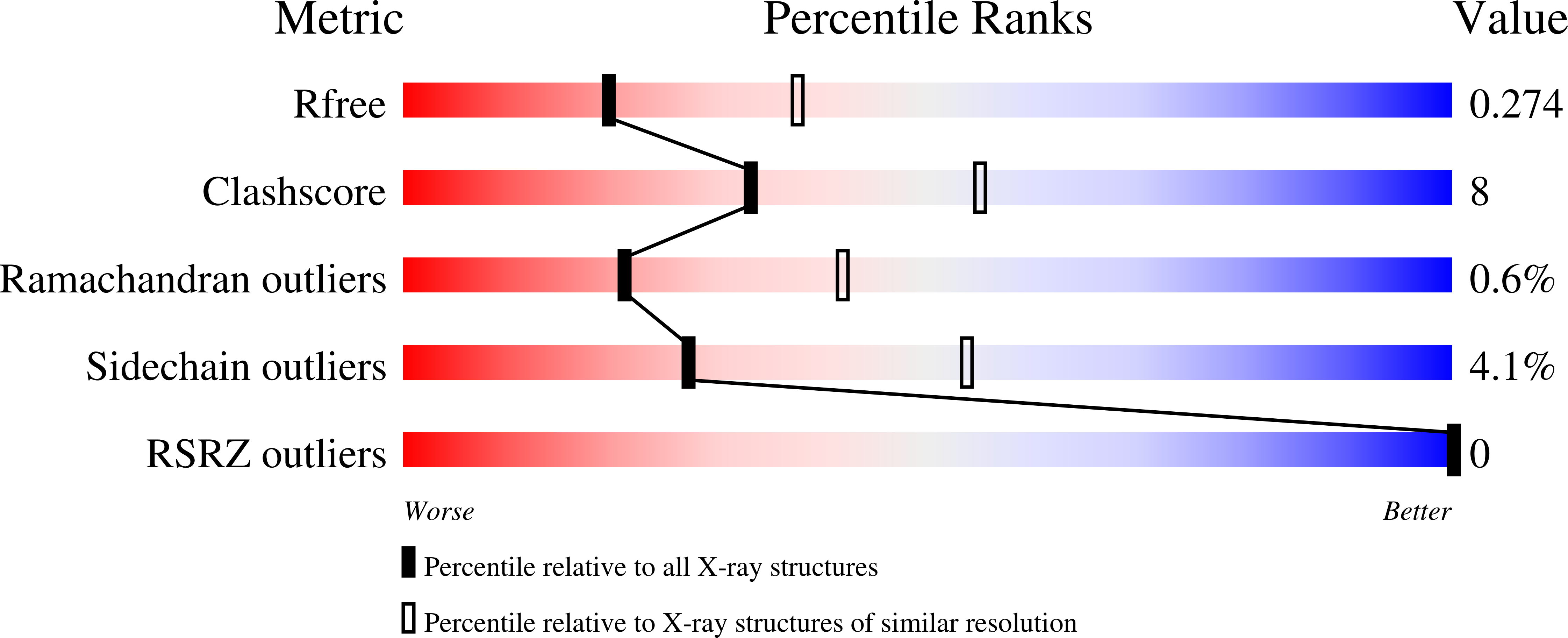
Experiments and Validation Details
wwPDB Validation report is not available for this entry.
X-ray source:
APS BEAMLINE 22-ID
Spacegroup:
P1
Expression system: Homo sapiens
