

ETHER</span>;</li> <li class='image_legend_li'>1 copy of <span class='highlight'>TRIETHYLENE GLYCOL</span>;</li> <li class='image_legend_li'>1 copy of <span class='highlight'>ACETATE ION</span>;</li> <li class='image_legend_li'>1 copy of <span class='highlight'>water</span>.</li></ul>")
ETHER</span>;</li> <li class='image_legend_li'>1 copy of <span class='highlight'>TRIETHYLENE GLYCOL</span>;</li> <li class='image_legend_li'>1 copy of <span class='highlight'>ACETATE ION</span>;</li> <li class='image_legend_li'>1 copy of <span class='highlight'>water</span>.</li></ul>")
ETHER</span>;</li> <li class='image_legend_li'>1 copy of <span class='highlight'>TRIETHYLENE GLYCOL</span>;</li> <li class='image_legend_li'>1 copy of <span class='highlight'>ACETATE ION</span>;</li> <li class='image_legend_li'>1 copy of <span class='highlight'>water</span>.</li></ul>")
Function and Biology Details
Biochemical function:
Biological process:
Cellular component:
Sequence domains:
- Ribosomal RNA adenine methyltransferase KsgA/Erm
- Ribosomal RNA adenine dimethylase
- S-adenosyl-L-methionine-dependent methyltransferase superfamily
- rRNA adenine dimethylase-like, C-terminal
- Ribosomal RNA adenine methylase transferase, conserved site
- Ribosomal RNA adenine methylase transferase, N-terminal
Structure analysis Details
Assembly composition:
monomeric (preferred)
Assembly name:
PDBe Complex ID:
PDB-CPX-279360 (preferred)
Entry contents:
1 distinct polypeptide molecule
Macromolecule:
Probable ribosomal RNA small subunit methyltransferase A
Molecule details ›
Chains: A, B
Length: 290 amino acids
Theoretical weight: 33.37 KDa
Source organism: Pyrococcus horikoshii OT3
Expression system: Escherichia coli BL21(DE3)
UniProt:
Sequence domains: Ribosomal RNA adenine dimethylase
Length: 290 amino acids
Theoretical weight: 33.37 KDa
Source organism: Pyrococcus horikoshii OT3
Expression system: Escherichia coli BL21(DE3)
UniProt:
- Canonical:
 O59487 (Residues: 1-268; Coverage: 100%)
Sequence domains: Ribosomal RNA adenine dimethylase
Ligands and Environments


Experiments and Validation Details
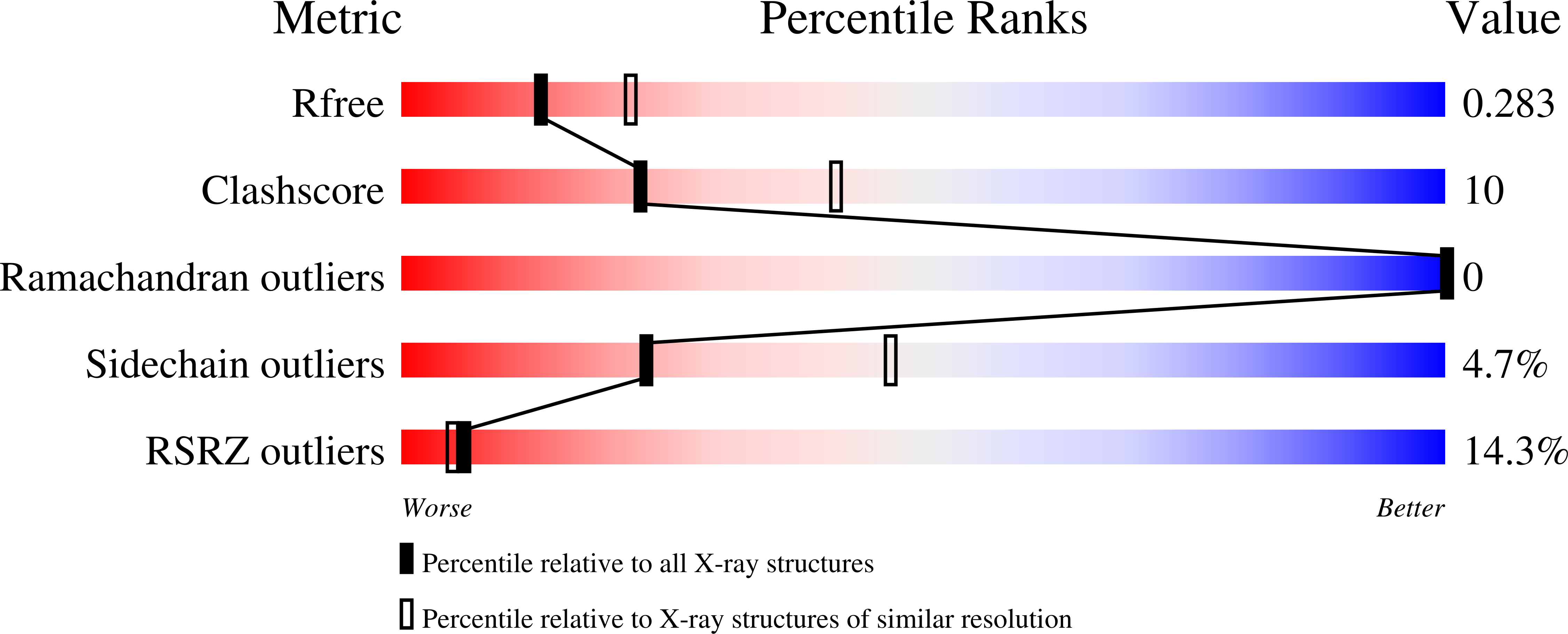
wwPDB Validation report is not available for this entry.
X-ray source:
RIGAKU MICROMAX-007 HF
Spacegroup:
C2
Expression system: Escherichia coli BL21(DE3)
